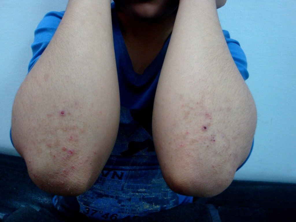

Diagnosis: Dermatitis herpetiformis
Description: Symetrical itchy excoriated vesicles
Clinical Features: Vesiculobullous
Pathology/Site Features: Elbow
Sex: M
Age: 18
Submitted By: Ian McColl
Differential DiagnosisHistory:
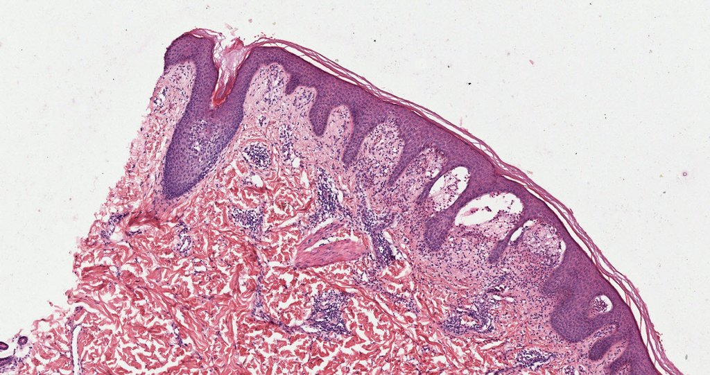
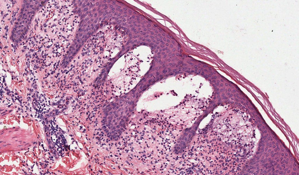
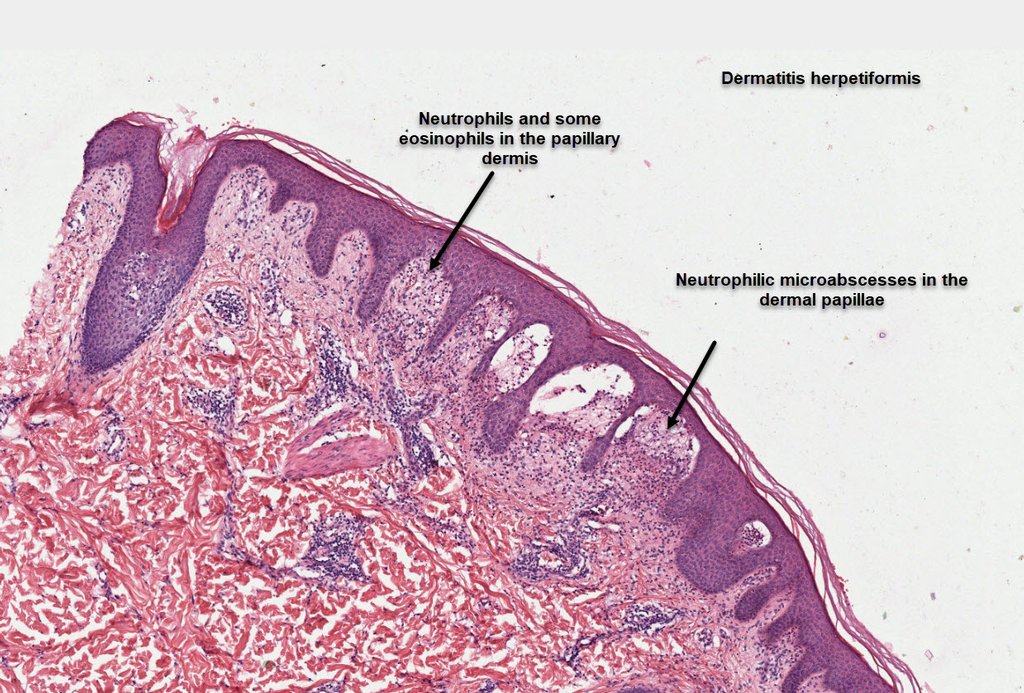
Histopathology of Dermatitis Herpetiformis Dermnet on Histopathology of Dermatitis Herpetiformis.
Clinical - Dermatitis herpetiformis presents often as crusts or vesicles over the extensor elbows, knees, buttocks, and across the scapulae, usually symmetrically, in someone with a history of coeliac disease. Because it is so itchy the vesicles and blisters are burst forming crusts. However many cases of DH are associated with non specific dermatitis like signs and blind biopsies may be needed with immuno.Also measure tissue transglutaminas and anti endomysial antibodies.
Histology - In H/E there are collections of neutrophils in the dermal papillae but the clincher is the finding of granular IgA in the dermal papillae associated with these neutrophils.(Vertical picket fence appearance). About a third of patients may have a bit of linear IgA deposition as well. Dont biopsy eroded lesional skin! Nuclear dust in the dermis is seen in 3 conditions , leukocytoclastic vasculitis, Sweets and Dermatitis herpetiformis.Initially there is fibrin with necrosis and acantholysis and small lacunae before a subepidermal vesicle forms.Eosinophils may be seen in older patients.
DDs - Linear IgA disease, Inflammatory type of EBA, Bullous SLE, Cicatricial pemphigoid and pustular vasculitis.
View this Clinical Case in GlobalSkinAtlas


