

Diagnosis: Tinea pedis
Description: Sub corneal vesicle
Clinical Features: Vesicles
Pathology/Site Features: Toes
Sex: M
Age: 35
Submitted By: Ian McColl
Differential Diagnosis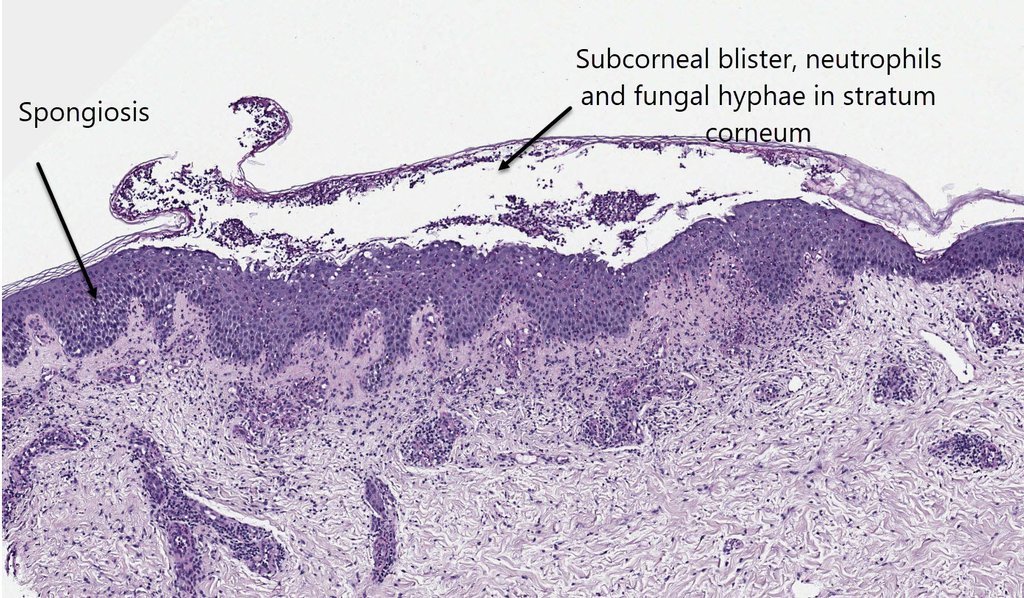

History:
Histopathology of Superficial Fungal Infections Dermnet on Tinea corporis
Clinical -A red scaly rash with clearing in the centre. Steroid modified tinea may lack the scale
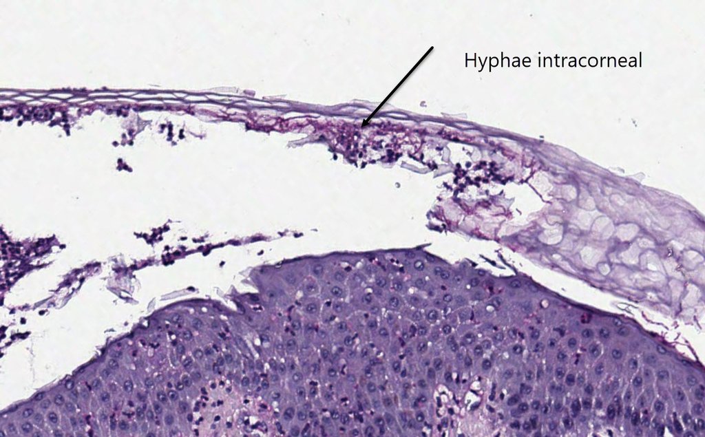
Histology- Basket weave keratin with fungal hyphae in the stratum corneum. In acute cases there may be neutrophils in the stratum corneum.
DD- Psoriasis with neutrophils in stratum corneum and also impetigo.
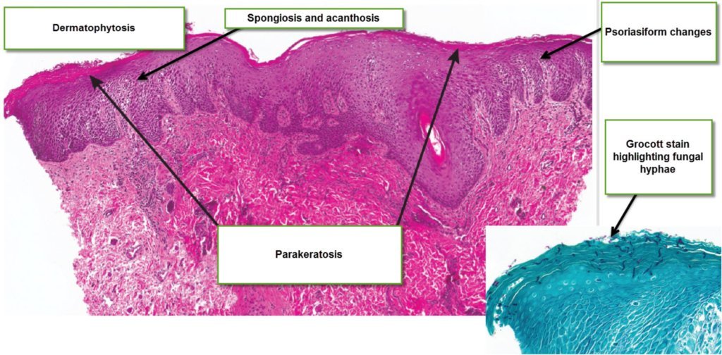
Dermatophytosis is the generic name given to the various clinical presentations of dermatophyte fungi. The fungal organisms are seen either in the keratin of the stratum corneum or on the outside (ectothrix) or inside (endothrix) of keratin hairs or in the keratin of the nails. The histology can be fairly bland or quite inflammatory depending on the immune reaction to the fungus and whether topical steroids have been applied to the rash.
Histology - Usually compact orthokertatosis but also the "sandwich" sign with orthokeratosis or parakeratosis alternating in layers with the basket weave stratum corneum. In more inflammatory cases neutrophils are seen in the stratum corneum. See Virtual Slide
Epidermis can be normal, show psoriasiform or spongiotic changes, subcorneal pustules like psoriasis and sometimes bullae. The SPI is a mixture of lymphocytes, eosinophils and neutrophils in some cases. View Virtual slide of Bullous dermatophyte infection
View this video of Bullous Tinea Pedis