
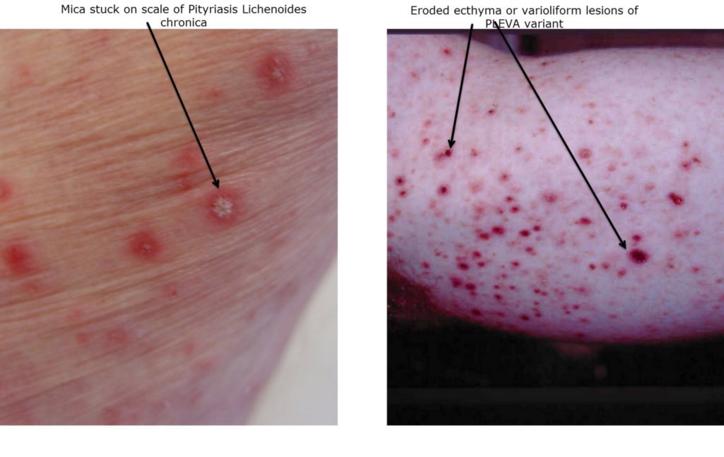
Diagnosis: Pityriasis lichenoides
Description: A mixture of papulonecrotic lesions and red scaly lesions
Clinical Features: Crusts and scales
Pathology/Site Features: Chest
Sex: M
Age: 23
Submitted By: Ian McColl
Differential Diagnosis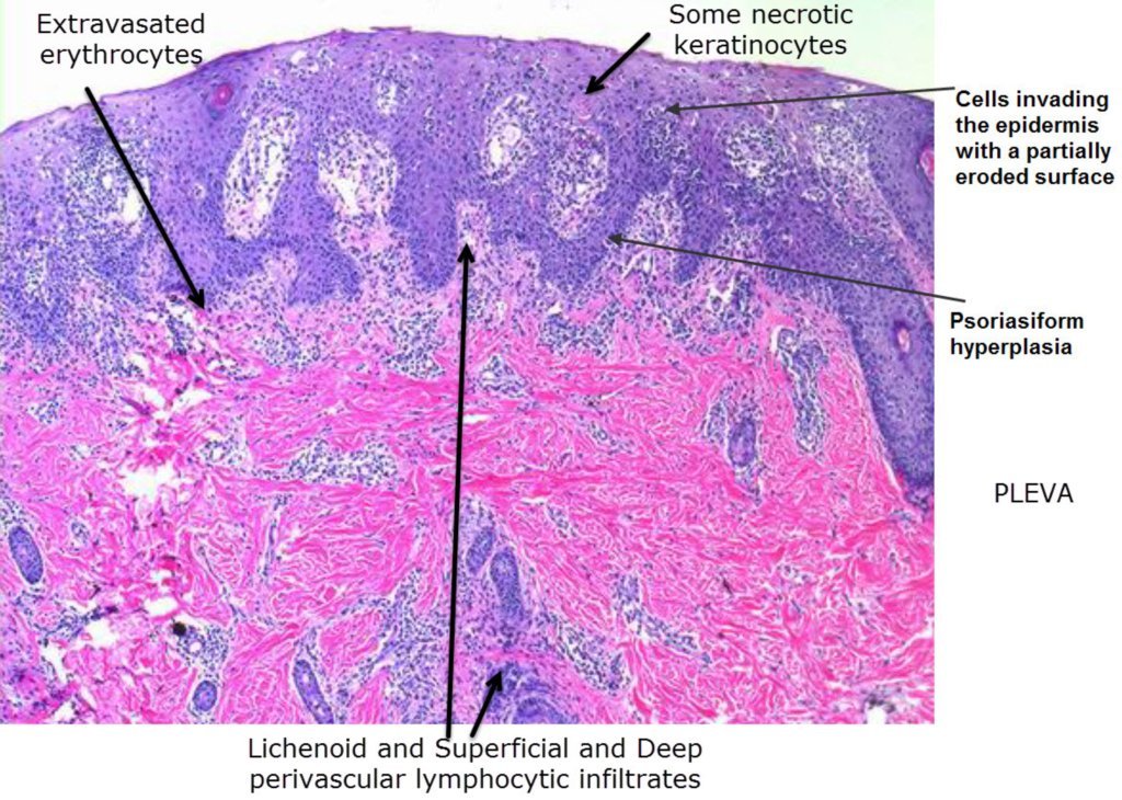
History:
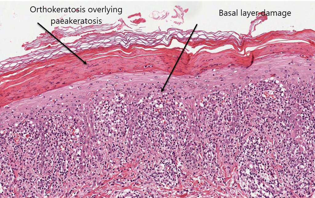
Pityriasis Lichenoides A red scaly disease with an acute and chronic presentation , Pityriasis lichenoides Chronica and PLEVA Dermnet on PLEVA
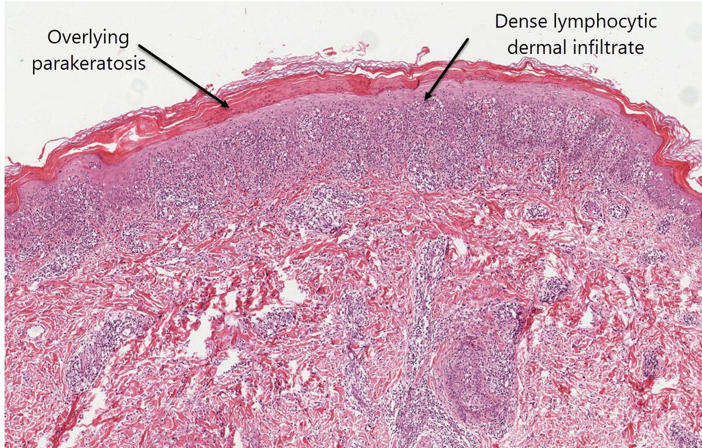
The lichenoid infiltrate is more prominent in PLEVA, especially the more acute form Virtual Slide where again there will be basal layer damage, but there is usually some degree of overlying epidermal thickening and parakeratosis corresponding to the scale that is seen in this disorder. There is also dyskeratosis of the keratinocytes as well and a characteristic feature of the dermal infiltrate of lymphocytes is it's wedge shaped distribution. There is a chronic form of this condition Virtual Slide where the dyskeratotic cells won't be as prominent, but there is still a degree of parakeratosis and the lichenoid infiltrate won't be as prominent as well. In late secondary syphilis virtual slide you can get quite a marked lichenoid pattern but this time as well as the lymphocytes you will see a lot of plasma cells and you should not see much in the way of eosinophils. View this video on the Clinical and Histological features of Pityriasis Lichenoides DX Path Pityriasis Lichenoides Chronica