

Diagnosis: Desmoplastic Melanoma
Description: Firm pink nodule back
Clinical Features: Nodule pink
Pathology/Site Features: Back
Sex: M
Age: 65
Submitted By: Ian McColl
Differential Diagnosis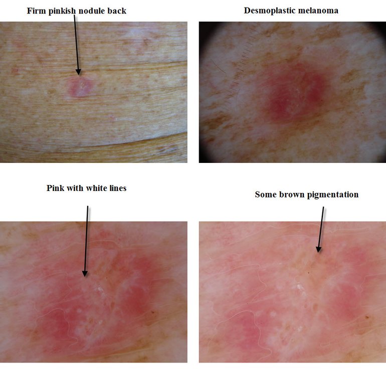
History:
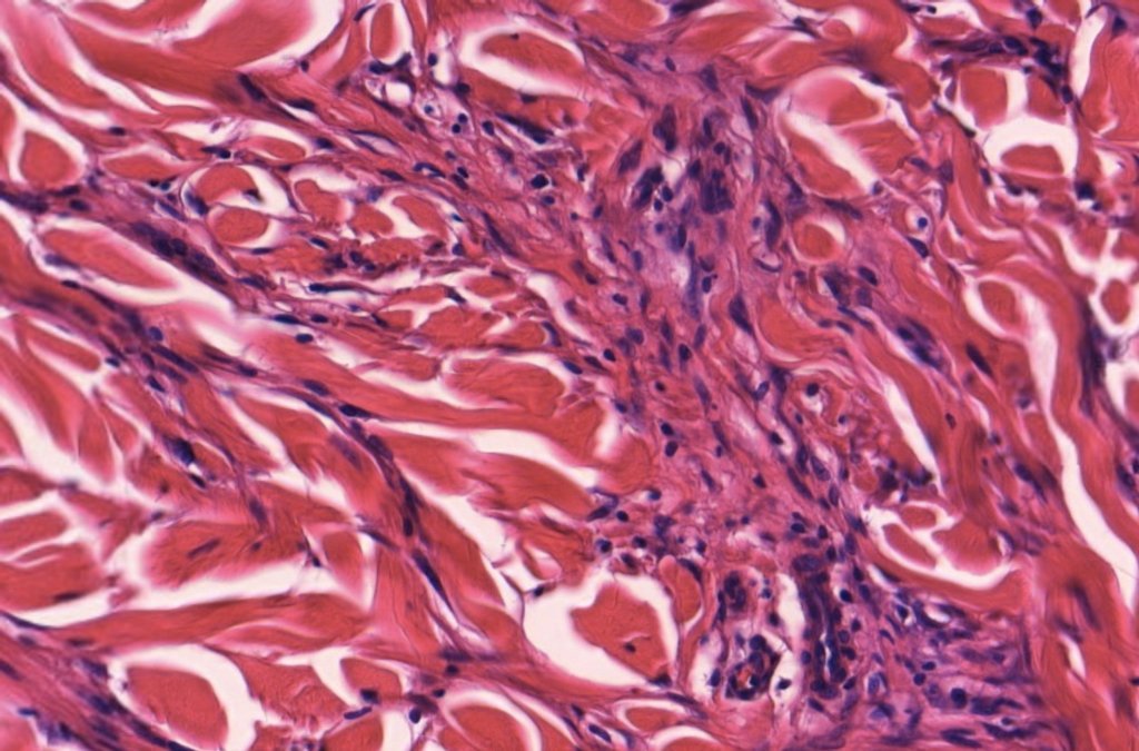
Desmoplastic Melanoma - Pink lesion on the back or one with lentigo maligna on the surface but a firm underlying base. Virtual Slide of Desmoplastic melanoma Virtual slide with Lentigo maligna
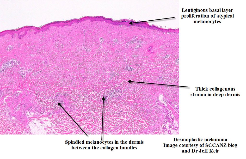
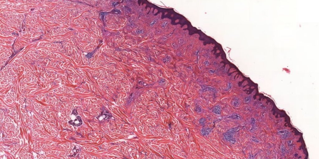
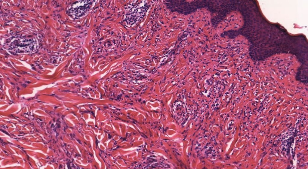
Histological Features Spindled cell tumour of dermis with marked surrounding fibrosis and collagen formation.There may be a Grenz zone but extends in depth often into fat. Lymphocytic accumulation as small nodules or follicle like areas are sometimes seen. Recurrent tumour can look like a scar and special stains are required. Some cases show an overlying epidermal lentigo maligna change.Sometimes the spindled cells have a whorled or storeyform arrangement simulating a dermatofibroma or DFSP.