
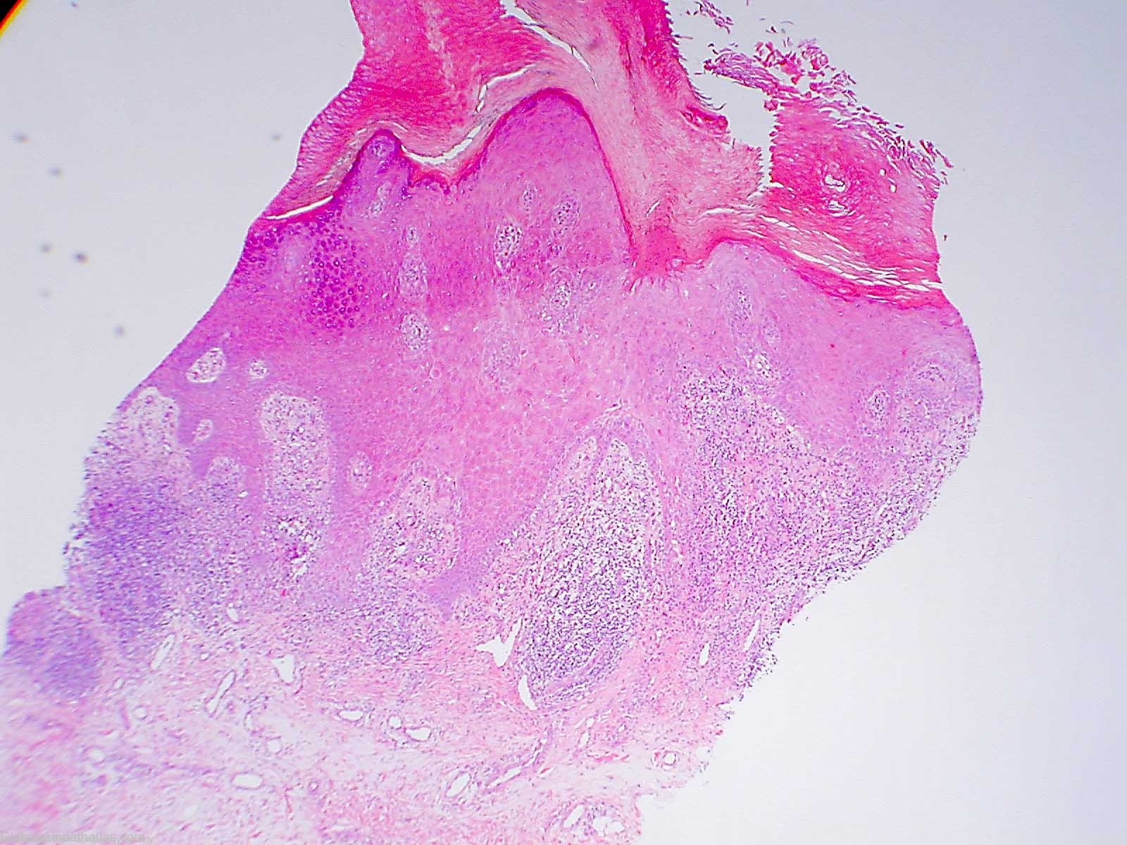
Diagnosis: Hypertrophic Lichen planus
Description: Thickened epidermis with hypergranulosis,sawtoothed rete ridges and lichenoid basal layer degeneration
Clinical Features: Papules
Pathology/Site Features: Pseudoepitheliomatous hyperplasia
Sex: M
Age: 62
Submitted By: Ian McColl
Differential Diagnosis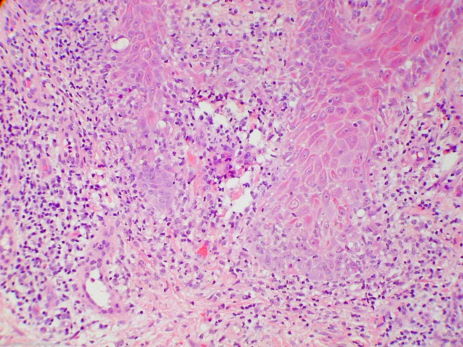
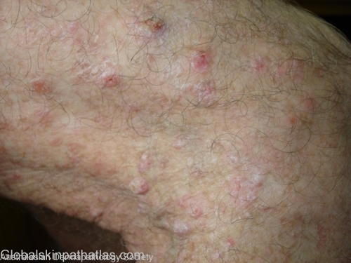
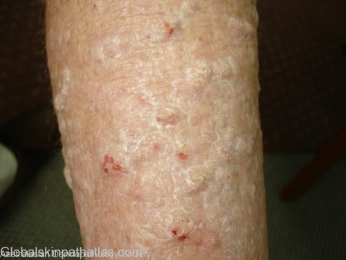
History:
Man in his late 60s with a three month history of these itchy keratotic papules on both lower legs.Biopsies have been taken from two of these lesions around the knee.He has had a strong topical steroid on them for 3 weeks.Exclude SCCs.They look like prurigo papules.
Comments: There is marked epidermal hyperplasia with lichenoid change at the tips of the rete ridges accompanied by upper dermal inflammation. These features most suggest hypertrophic lichen planus.
See the recent article by Kossard in Archives of Dermatology