

Diagnosis: Mycosis fungoides
Description: Erythematous scaly plaques
Clinical Features: Plaque
Pathology/Site Features: Buttocks
Sex: M
Age: 49
Submitted By:
Differential Diagnosis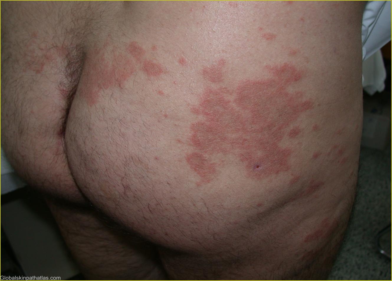
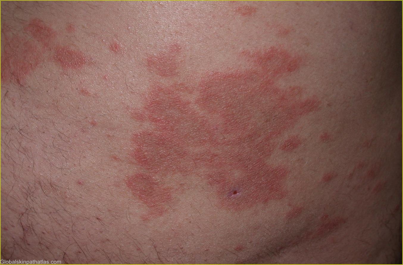
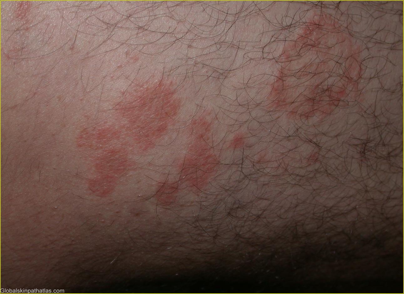
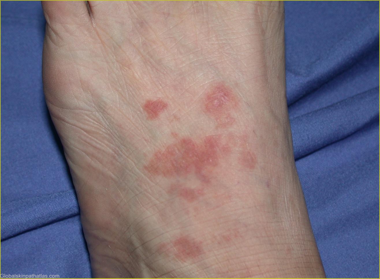
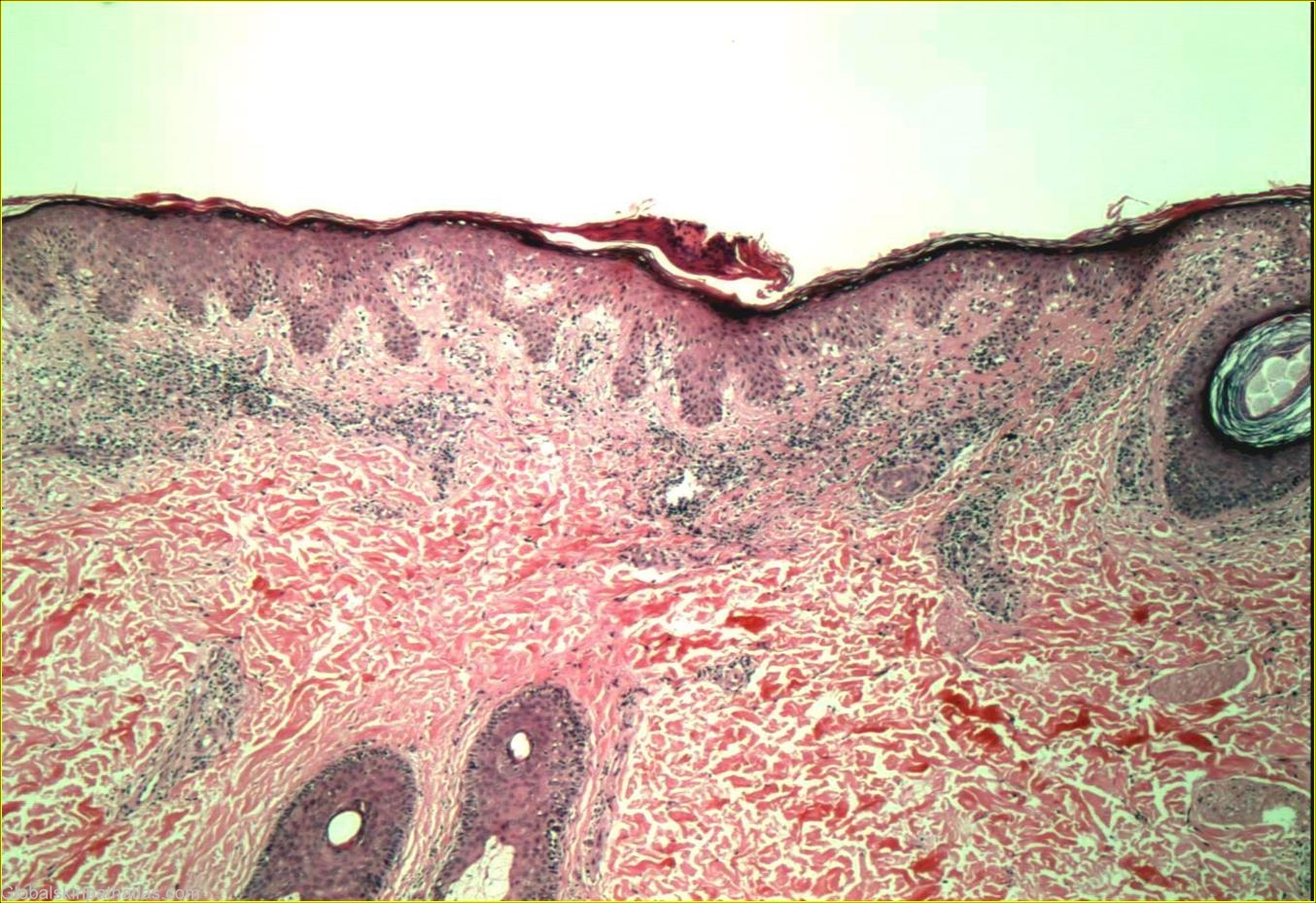
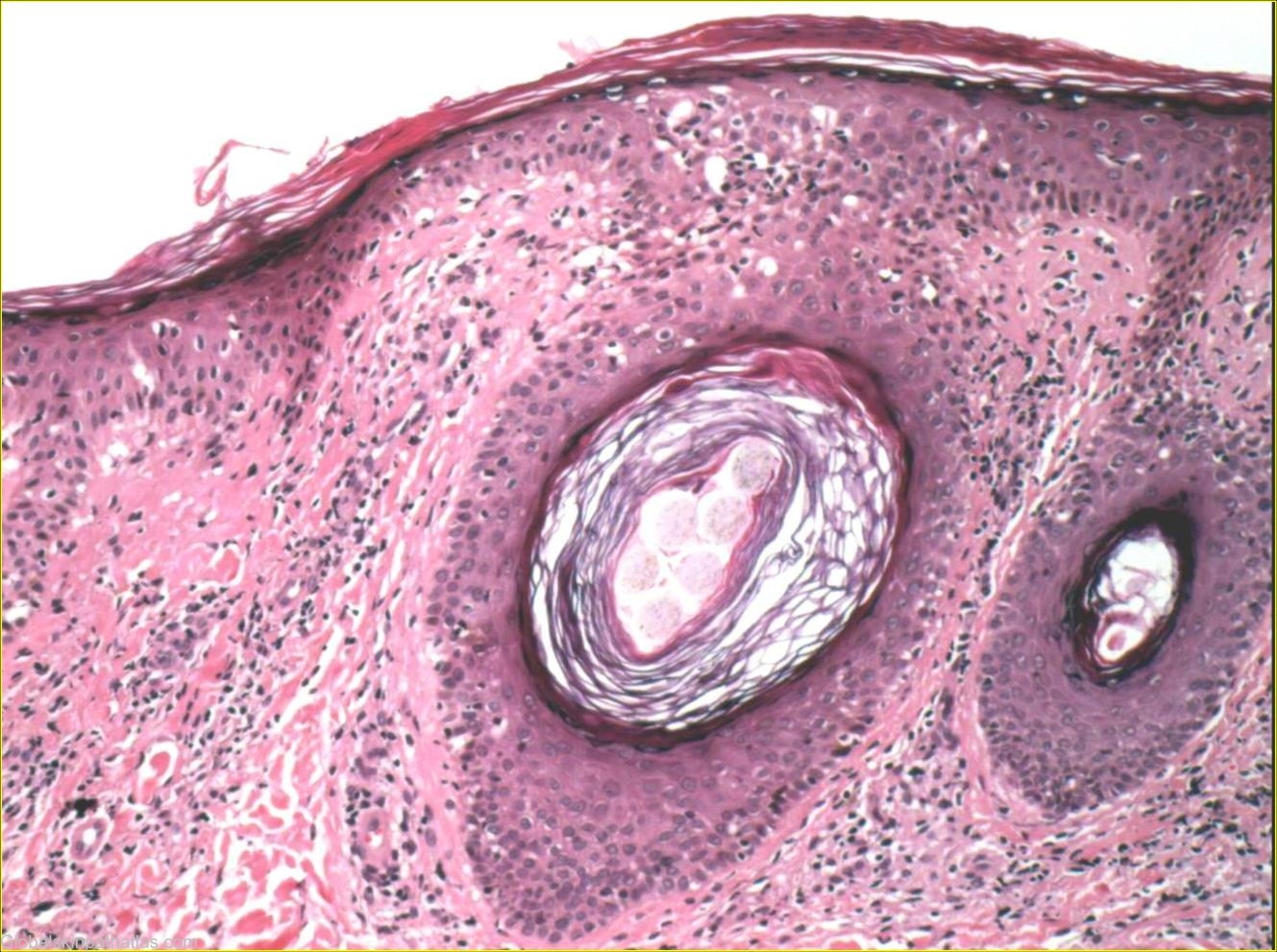
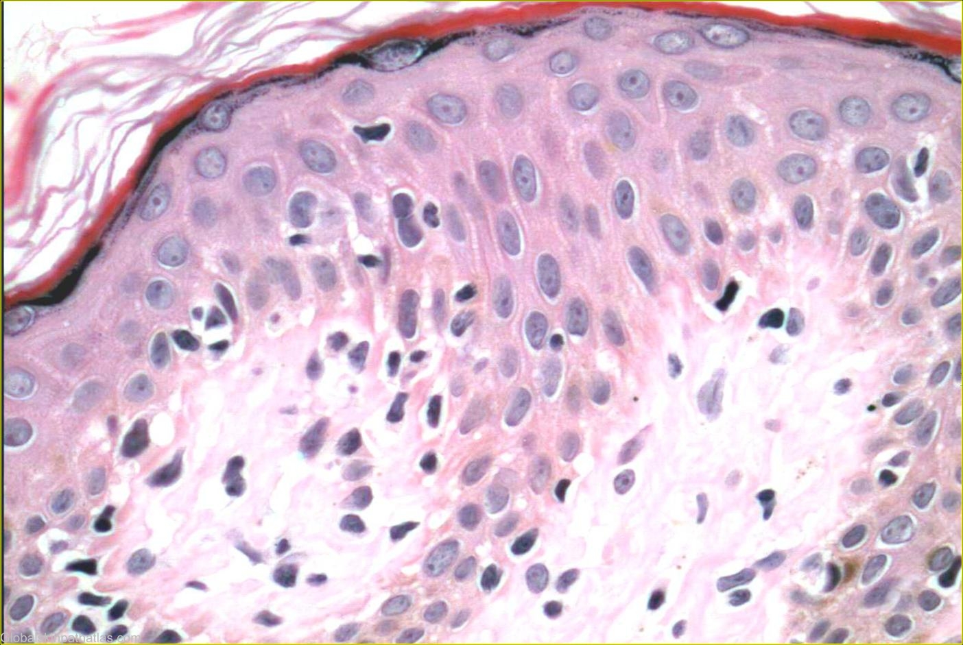
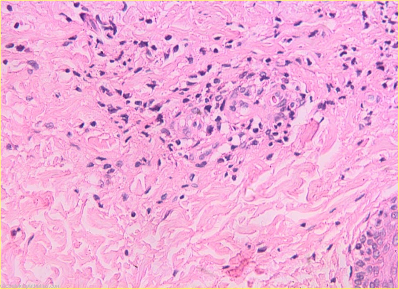
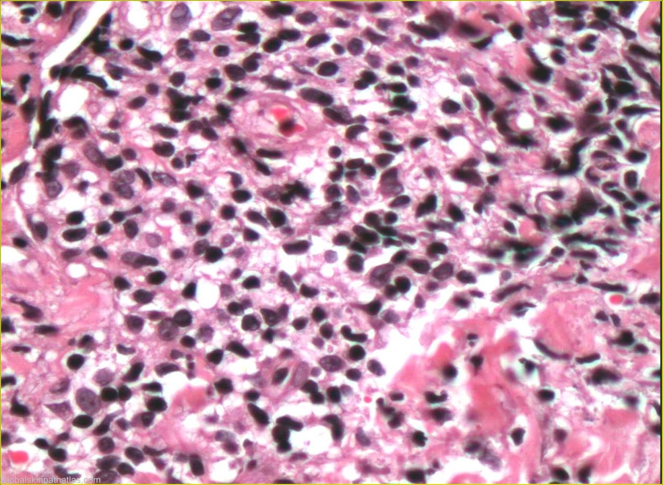
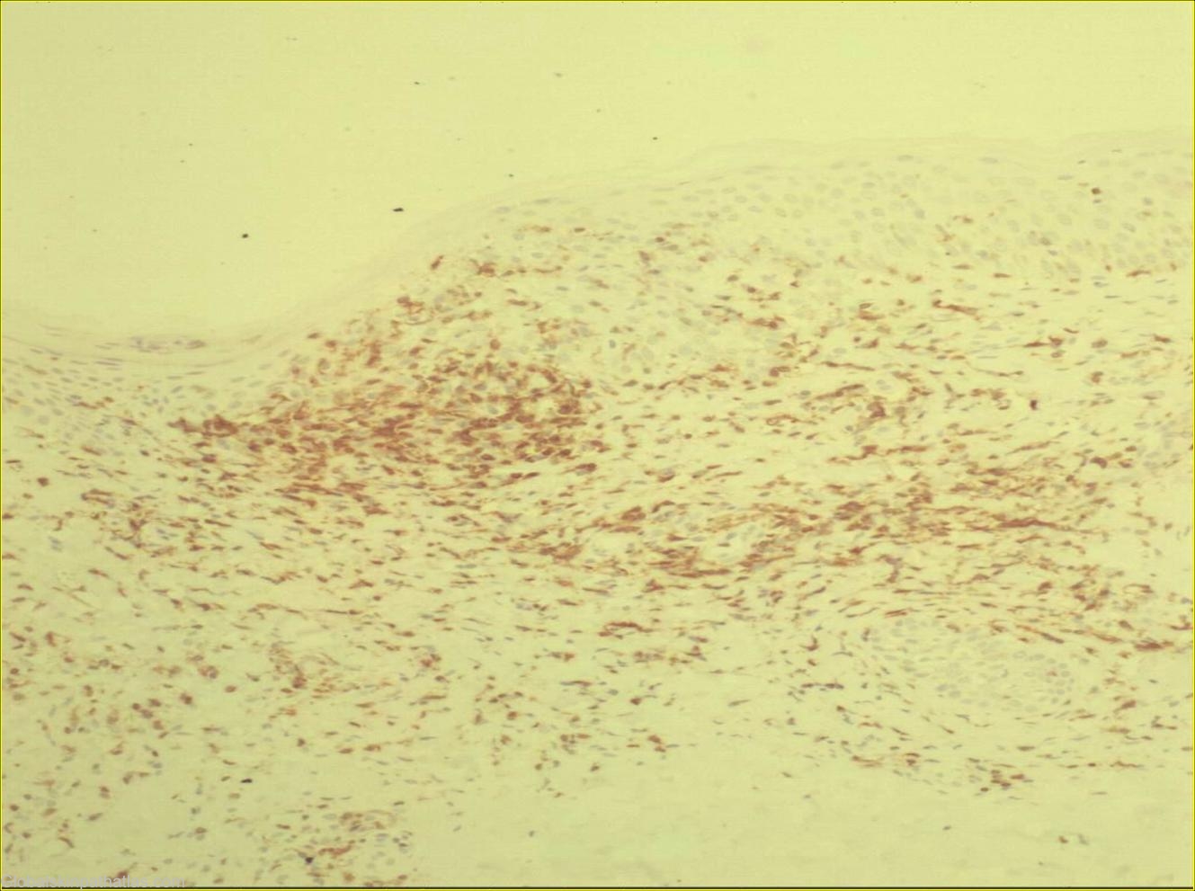
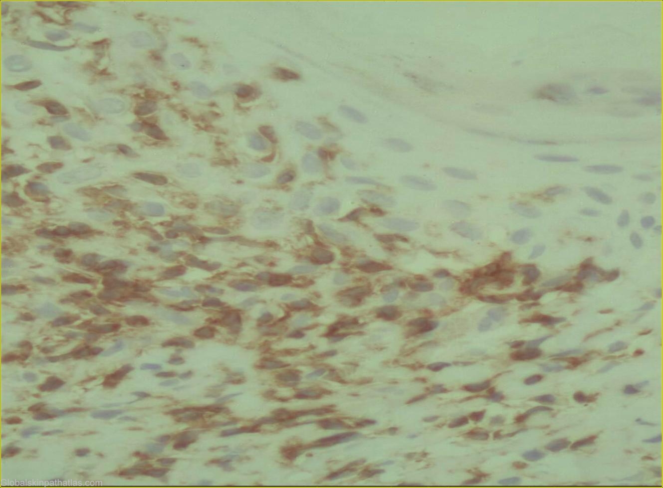
History: This 49-year-old male presented with bilateral non-itchy erythematous scaly plaques of two months duration mainly on buttocks, sacrum, thighs and feet. No history of drug intake and no history of similar lesions in family members. Sections show a biopsy of skin in which the epidermis shows psoriasifrom hyperplasia with dense superficial and band-like dermal infiltrate. Focal liquefaction degeneration of the basal layer as well as abnormal lymphocytes both singly and in groups are also evident in the epidermis. The abnormal lymphocytes are also present in the dermis. These cells stain positively for CD3 and CD4.