
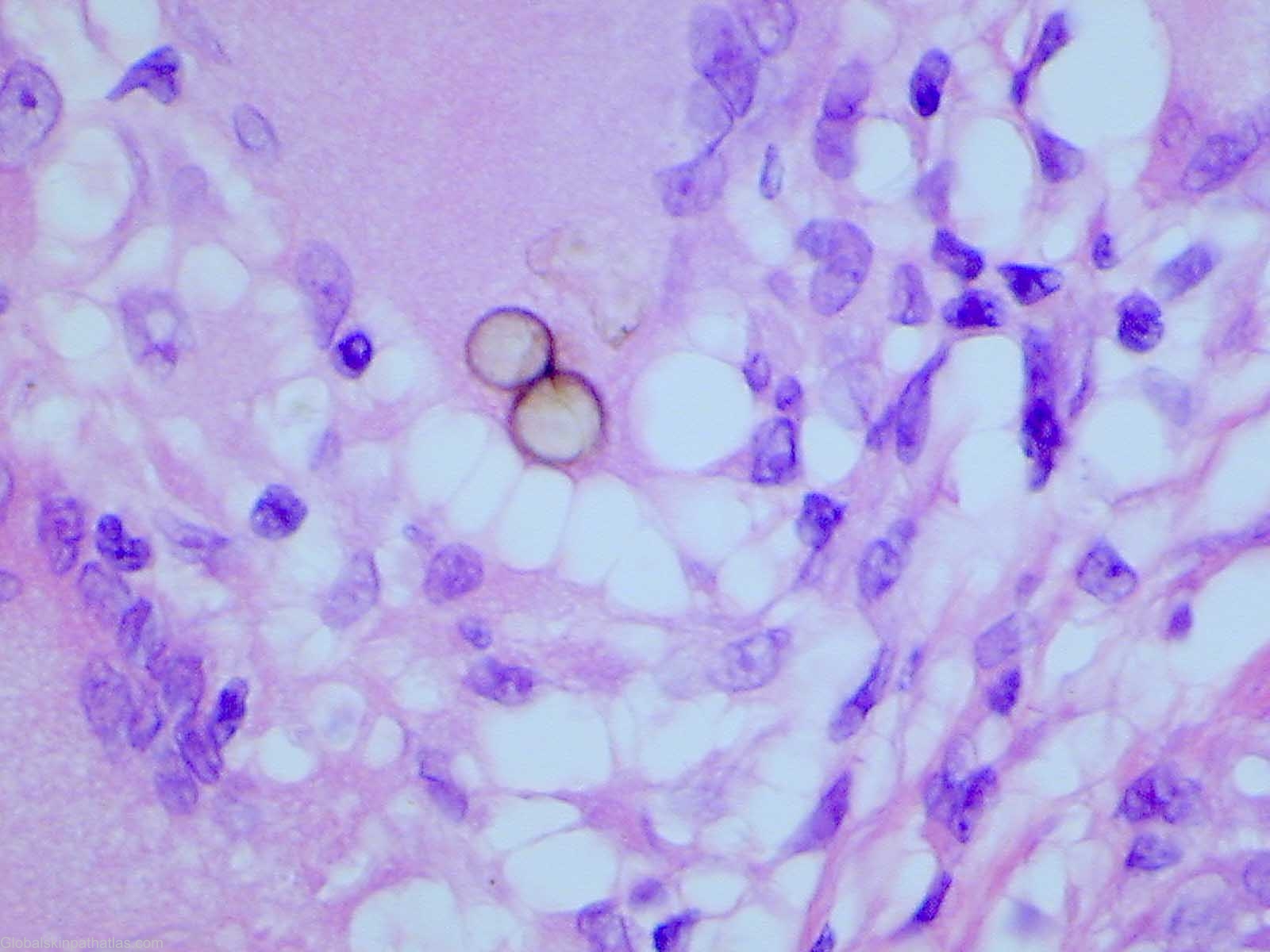
Diagnosis: Chromoblastomycosis
Description: Copper pennies in the dermis fungal spores
Clinical Features: Papules,red
Pathology/Site Features: Brown black in dermis
Sex: M
Age: 45
Submitted By: Ian McColl
Differential Diagnosis
History:
This lesion was a small pink papule on a man's wrist which was excised as a presumptive BCC.It was barely 3mm in diameter.He had no other lesions.
The histopathology usually shows pseudoepitheliomatos hyperplasia with intraepidermal microabscesses.There is usually a mixed dermal infiltrate including plasma cells and histiocytes with multinucleated giant cells but no caseation.The "copper pennies" are clusters or chains of brown spores,sometimes within histiocytes and also free in the tissues.
The pathology report was as follows
Sections show a granulomatous reaction involving the upper and mid dermis. The granulomas are formed by rather tight collections of mono-nucleated and multi-nucleated giant cells. A moderate infiltrate of lymphocytes surrounds the granulomas. Several of the granulomas contain large brown yeast organisms. Some of these are septate. These have the morphology of chromomycosis. There is no evidence of malignancy. Excision appears complete.