

Diagnosis: Acral lentiginous melanoma
Description: Atypical melanocytes ascending from the rete ridges
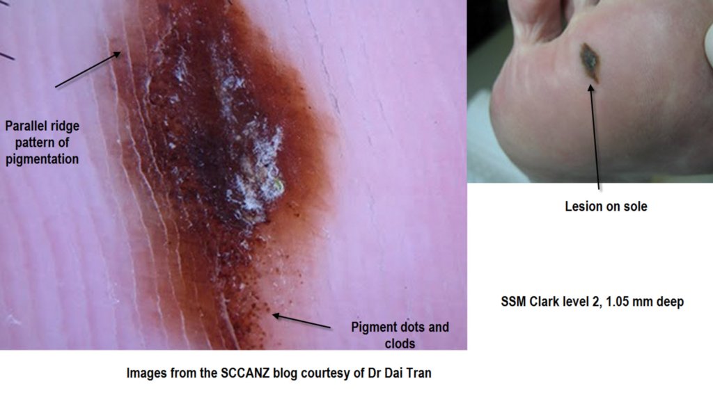
Clinical Features: Macule brown
Pathology/Site Features: Foot,sole
Sex: M
Age: 54
Submitted By: Ian McColl
Differential Diagnosis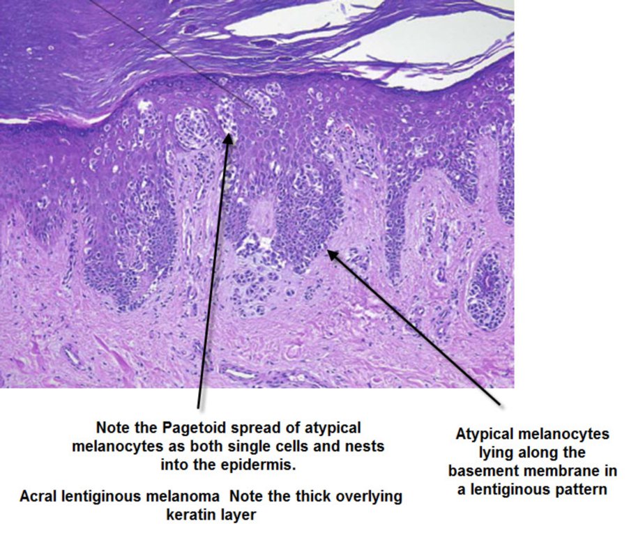
History:
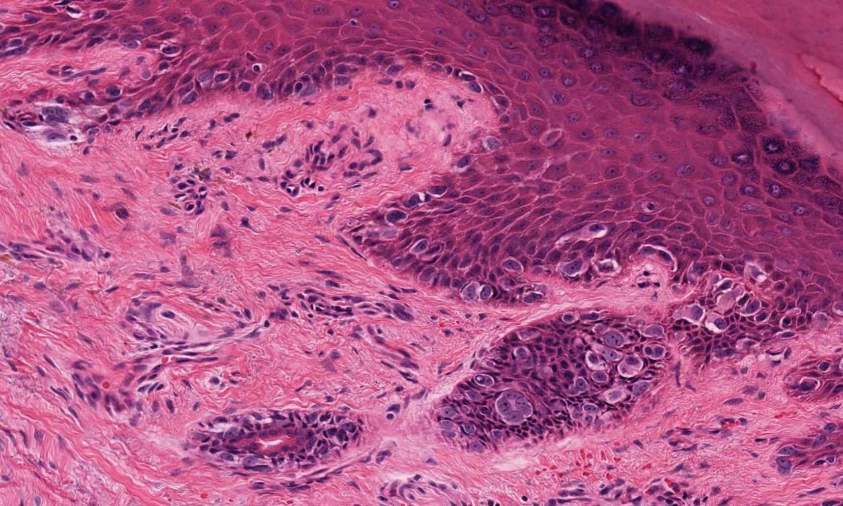
Histopathology and Dermatoscopy of Nevi of special sites particularly acral and genital
Junctional or compound melanocytic nevi on the skin of the palms, soles,nailbeds, knees and elbows feature upward scatter of melanocytes as often than not .
Upward migration of melanocytes can be found even in relatively small, clinically banal nevi at these sites. (Le Boit)
Cutaneous melanocytic nevi of so-called special sites (including the skin of ears, axillae, milk line, umbilicus, inguinal region, knee, elbow, wrist, ankle, acral skin), tend to exhibit more architectural and cytologic variability than those of other body sites. (Mooi)
Melanocytic nevi on or near genital skin have often been mistaken for melanoma
because they demonstrate lateral confluence of junctional nests, and some of the cells
that comprise those nests can ascend into the spinous and cornified layers.
Additionally, some nevi on or near genital skin have dense lymphocytic infiltrates in
the papillary dermis (Le Boit)
Look at this virtual slide from Path Presenter
Although occasional atypical naevi from the perineum are dysplastic, others fall into a category of atypical genital-type melanocytic naevus. They are most often seen in female patients—usually young women—but they are sometimes seen in children. They are characterised by a warty architecture. Large junctional nests surrounded by a well developed retraction artifact are a diagnostic clue. They may exhibit pronounced cytological atypia and occasional dermal mitoses, but do not display the architecture of a dysplastic naevus. Although dermal fibrosis is often a feature of these lesions, eosinophilic and lamellar fibroplasia is lacking.
Similar lesions may be encountered at other flexural sites, including the umbilicus, groin, submammary region, and axillae—hence their alternative names of flexural naevi and milk line naevi.
The biological potential of these worrying lesions is poorly documented and their histology is often alarming. It is important therefore to take note that vulval melanoma is very much a disease of the elderly and that these atypical naevi most often occur in the young.(Culpepper McKee)