
Diagnosis: Acne
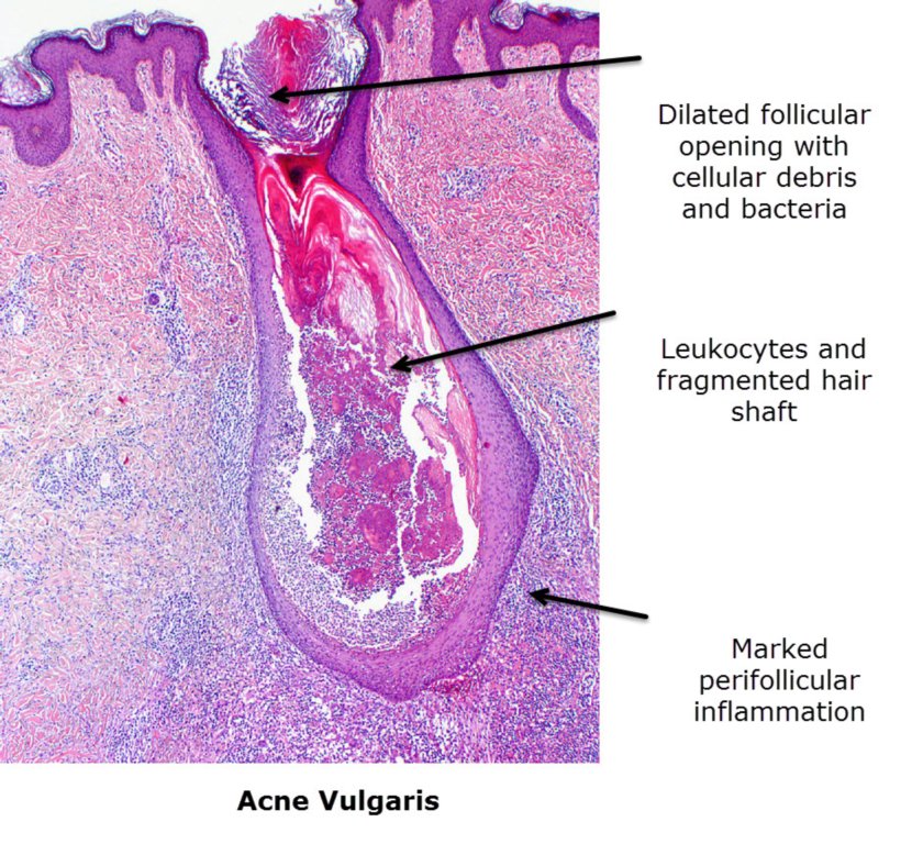
Description: histopathology of an acne pustule
Clinical Features: Pustules
Pathology/Site Features: Face
Sex: M
Age: 22
Submitted By: Ian McColl
Differential Diagnosis
History:
Acne is rarely biopsied. The histology depends on whether you biopsy a pustule or cyst. The histology below is of an acne pustule. A comedone will show laminated keratin in the follicular infundibulum.Rupture of a comedone releases keratin into the dermis inducing an inflammatory reaction and sometimes granuloma formation.
There is early follicular dilatation with retained keratin in layers in this histology image.. This is infiltrated by neutrophils. There is marked perifollicular inflammation. If the obstructed follicle ruptures there is a foreign body giant cell reaction. Open comedones or blackheads have large follicular openings while closed comedones have small openings. The DD include folliculitis where the inflammation is within the follicle and there are no comedones, Hidradenitis suppurativa with marked inflammation and sinuses and deep fungal or mycobacterial infections with organisms within granulomas.