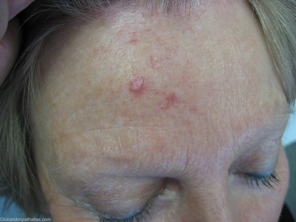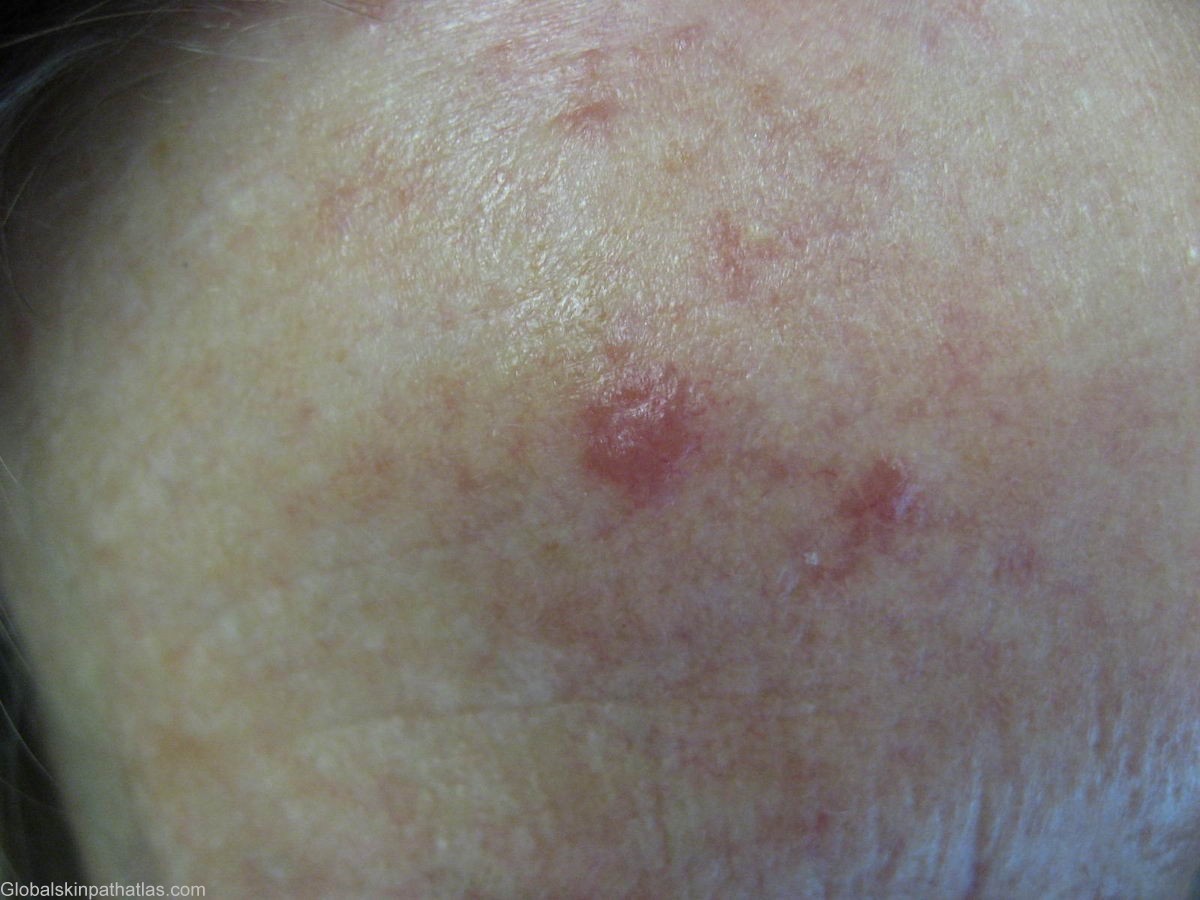

Diagnosis: Lymphoma Large B-cell
Description: pink nodules
Clinical Features: Nodule pink
Pathology/Site Features: Forehead
Sex: F
Age: 68
Submitted By: Ian McColl
Differential DiagnosisHistory: Elderly lady with a two month history of these two pink nodules on her forehead. Nothing elsewhere. They were asymptomatic.
DD of pink nodules The DD is very wide. A solitary pink nodule can be a tumour such as a BCC , malignant melanoma or even a pink seborrhoeic keratosis, a granulomatous process such as sarcoid or granuloma faciale, an infective lesion such as leishmaniasis and rare conditions such as Sweet's syndrome. Generally a biopsy is necessary to establish the diagnosis of the facial nodule and the tissue should be sent for culture for atypical mycobacteria and for deep fungal infections if an infective cause is considered.
Biopsies of these lesions were reported as Large B cell non Hodgkins lymphoma. She has no glands in her axillae , groin or neck. Scans have been done to see if there are any internal glands.Primary cutaneous B cell lymphoma may present as solitary or multiple cutaneous lesions. The treatment of choice is radiation therapy.
Some PCBCL can present as a feature of Borrelia infection as Lyme disease.
Primary cutaneous B cell lymphomas generally have a good prognosis with a 95% 5 year survival. They make up about 25% of all cutaneous lymphomas.
A Follicle centre lymphoma or Crosti's lymphoma is a solitary indolent nodule on the scalp. It is B cell and does not have any underlying nodal disease. Again radiotherapy is the treatment of choice.
Large B cell lymphoma is derived from germinal and post germinal centre cells and usually presents as a primary nodal lymphoma. They often are found on the lower limbs. The prognosis is poorer with a 5 year survival of 55%.
Follow Up
This lady appears to have a primary cutaneous B cell lymphoma. It was once thought all cutaneous B cell lymphomas represented cutaneous spread from an underlying nodal lymphoma.It has also been appreciated that many so called pseudolymphomas are in fact low grade malignant B cell lymphomas of the skin.
There are variations in the relative incidences of the 3 types of primary cutaneous B cell lymphomas in different countries and between continents. This may be due to different etiologies eg Borrelia in the environment. In Austria where Borrelia is endemic, Borrelia DNA sequences were found in 18% of cases of primary cutaneous B cell lymphoma.
All patients with a suspected primary cutaneous B cell lymphoma should still have a full workup including , complete blood examination,bone marrow biopsy and CT scans of the chest, abdomen and pelvis.Flow cytometry of peripheral blood and bone marrow cells can also be useful.
The three main subtypes of primary cutaneous B cell lymphoma are 1. Cutaneous follicle centre lymphoma, 2. Cutaneous marginal zone lymphoma and 3. Large B cell lymphoma (sometimes of the leg).
This lady's full path report was as follows.
The changes are similar in both biopsies and are suggestive of a cutaneous lymphoproliferative process. Immunohistochemical stains have been performed. CD20 (B cell marker) highlights the large atypical cells in both biopsies. A significant proportion of the cells are Bcl-2 positive. Small T cell lymphocytes (CD3 positive) are noted in the background. This profile is in keeping with a cutaneous B cell lymphoma.

