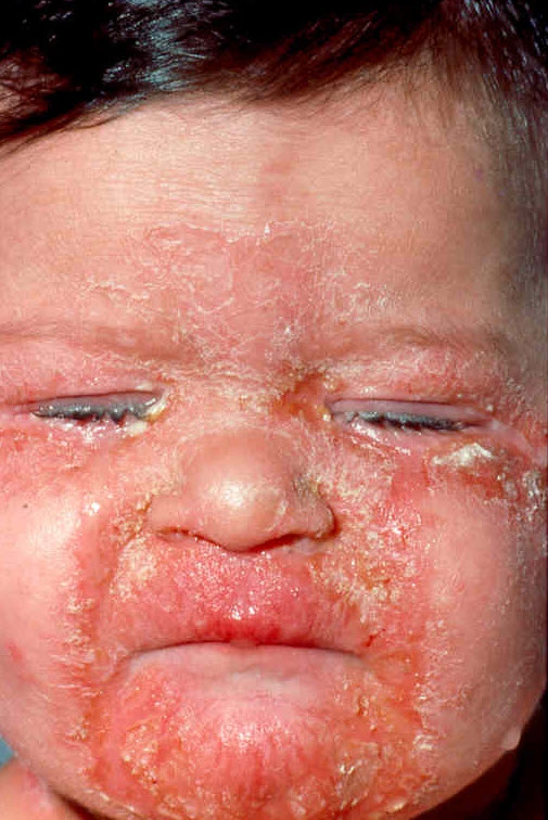
Diagnosis: Acrodermatitis enteropathica
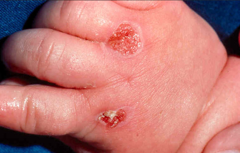
Description: Perioral erosions and crusts Sometimes this becomes impetiginised which can cause subcorneal neutrophils in the histology simulating psoriasis See the case in Global Skin Atlas
Clinical Features: Blisters
Pathology/Site Features: Face
Sex: M
Age: 1
Submitted By: Ian McColl
Differential DiagnosisHistory:
Acrodermatitis enteropathica is a rare disorder seen in infants when they are weaned or in premature infants with poor zinc stores.The clinical lesions look dermatitic with oozing and crusting particularly around the mouth,in the groins and acrally on the fingers and toes.Some lesions can be pustular or bullous.Chronic lesions can look more psoriasiform. The condition can also occur in adults with alcoholism,malabsorption or bowel inflammatory disease.
The condition rapidly resolves with oral zinc supplementation.Particularly be mindful of this condition in an infant with chronic diarrhoea and nappy rash especially if they develope a perioral rash.
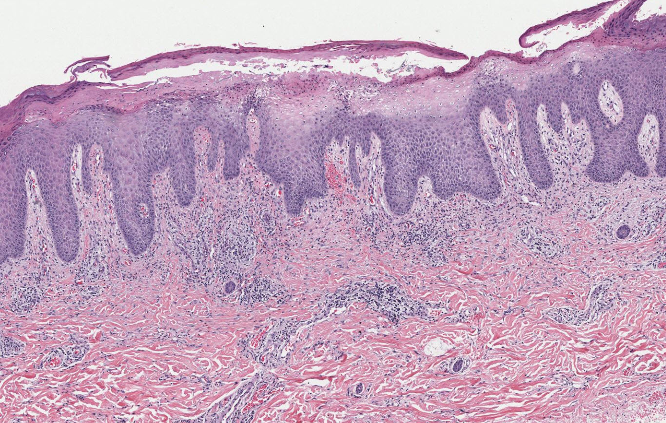
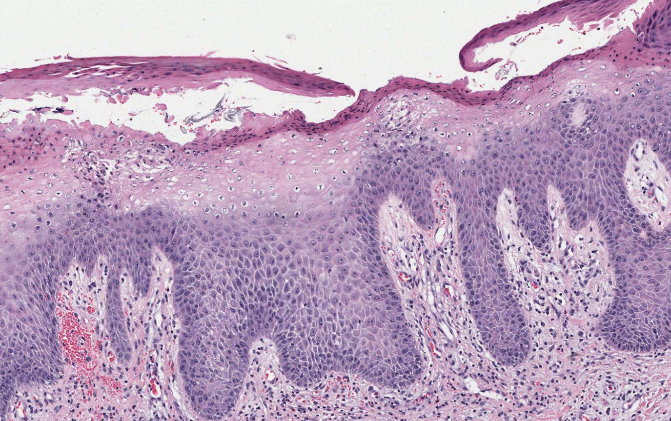
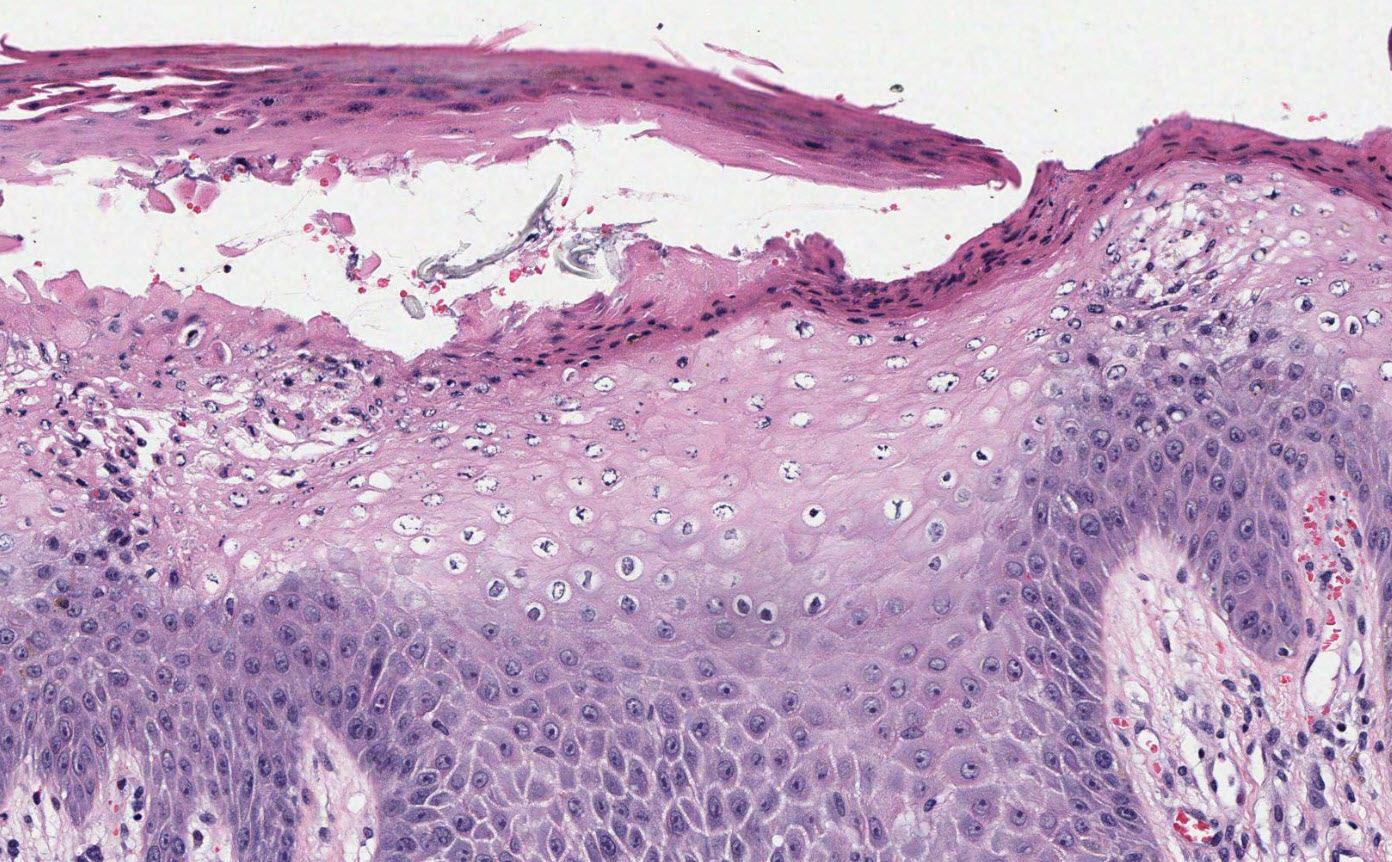
Histology Palor and or vacuolization of the upper epidermis with often confluent parakeratosis and loss of the granular layer. There may be psoriasiform hyperplasia making it look like psoriasis and the presence of neutrophils in intraepidermal vesicles may re inforce that thought. Subcorneal pustules do favour psoriasis but the palor and vacuolization are the stand out features of acrodermatitis enteropathica. Other conditions such as pellagra, necrolytic acral erythema, glucagonoma syndrome and biotin deficiency are also possible DDs that require clinical correlation to diagnose them acurately.
Path images courtesy of Path Presenter
