
Diagnosis: Nevus Congenital
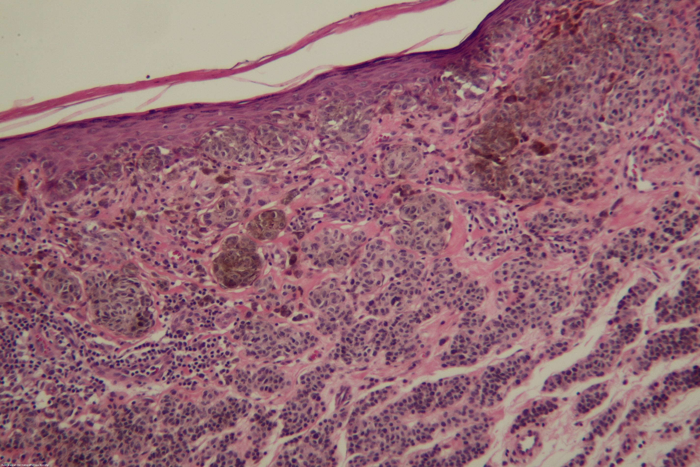
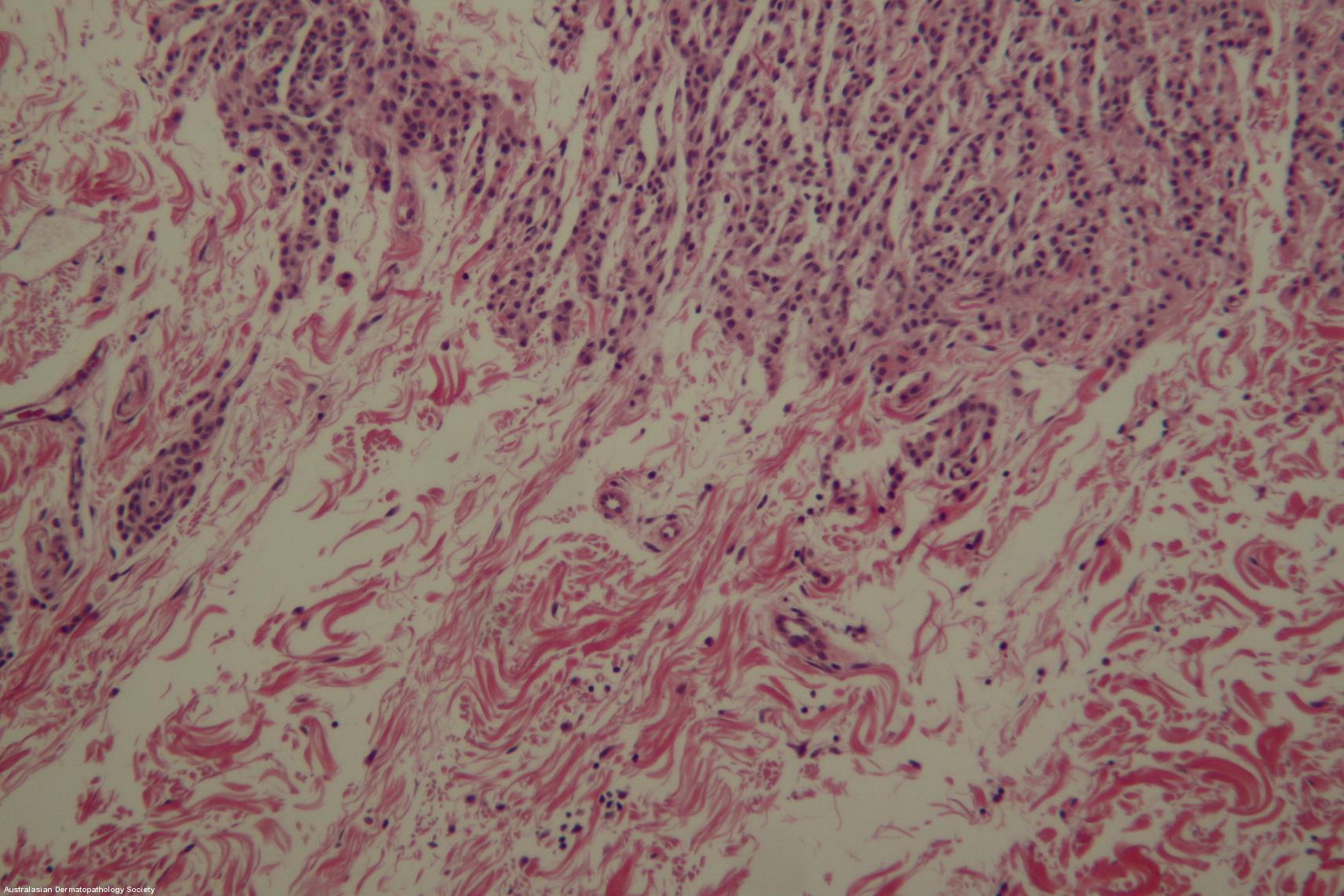
Description: Symmetrical compound melanocytic lesion with prominent junctional nests of melanocytes with focal bridging
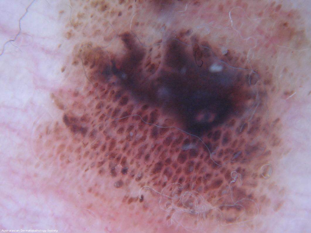
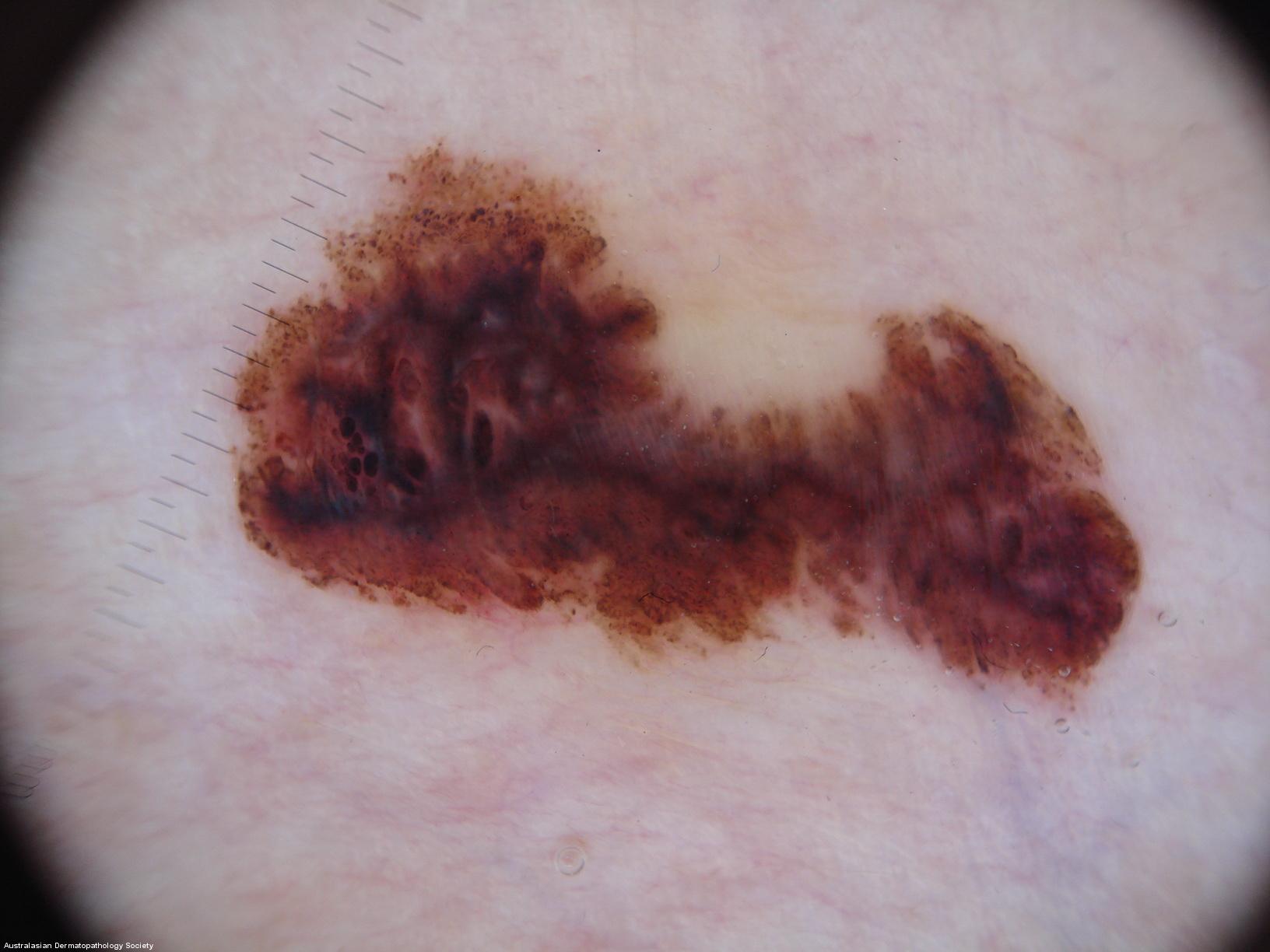
Clinical Features: Papules,black
Pathology/Site Features: Nevus cells dermis
Sex: M
Age: 15
Submitted By: Ian McColl
Differential Diagnosis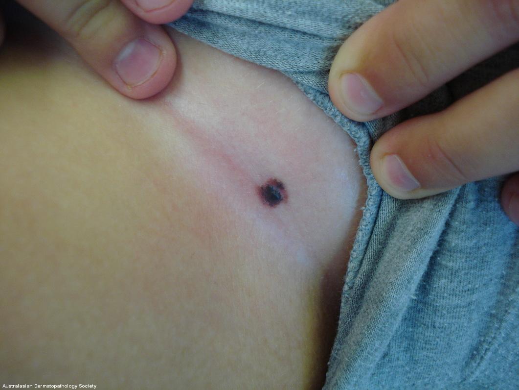

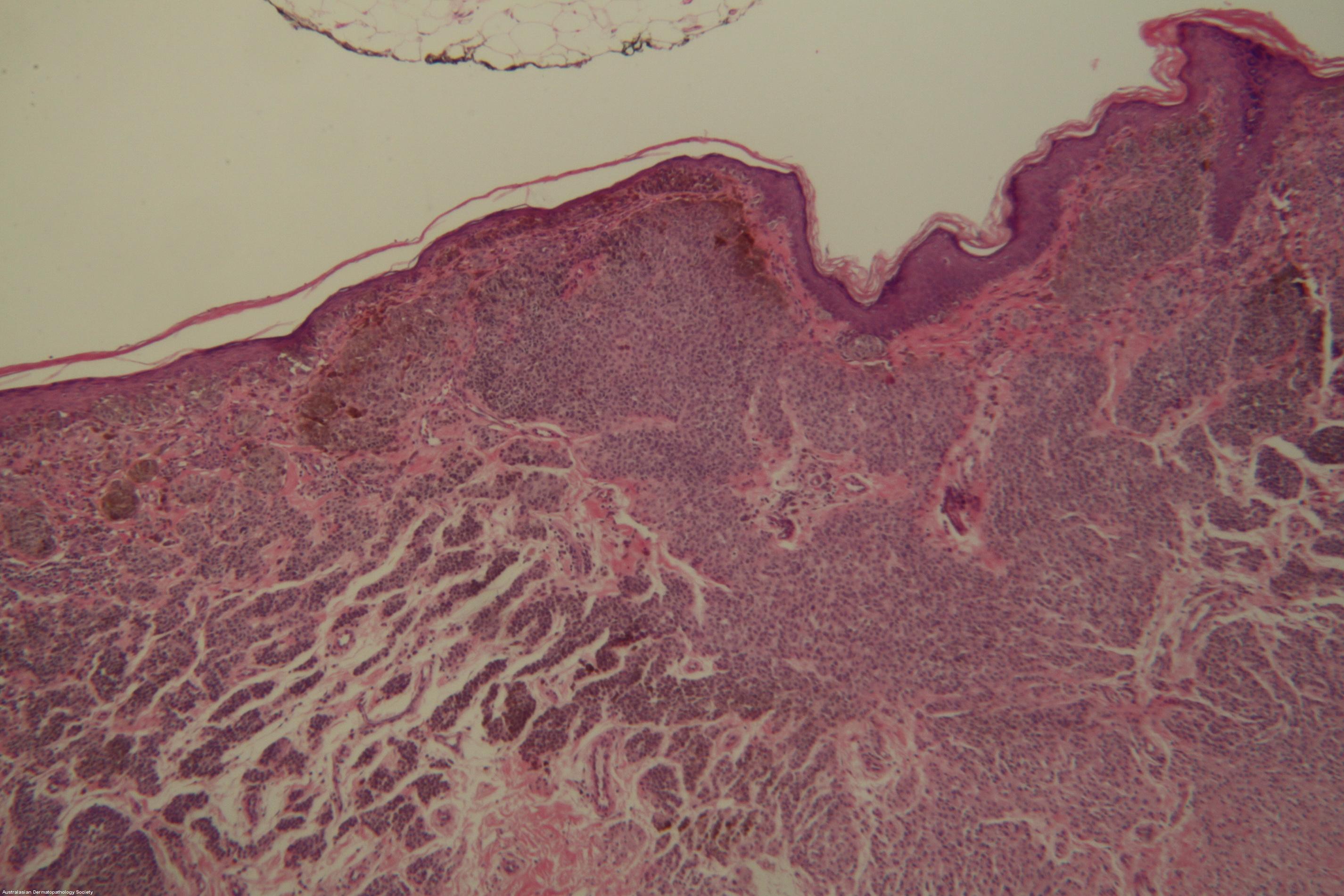
History: Initial lesion in groin was biopsied aged 9 and reported as compound nevus. Six years later he returned with further growth of lesion. Fully excised. Clinically a congenital nevus but not present at birth or first 3 years of life.
Description: No deep mitoses
Comments: Symmetrical compound melanocytic lesion with prominent junctional nests of melanocytes with focal bridging. No Pagetoid spread of single cells to upper epidermis. The dermal component matures in depth. Focally brisk lymphocytes and scattered melanophages. Appearances favour a dysplastic compound nevus with moderate to severe atypia. This is also a special site and nevi here may appear histologically worse than they actually are.