
Diagnosis: Lymphomatoid papulosis
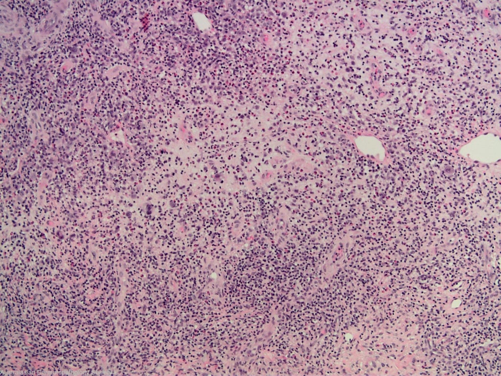
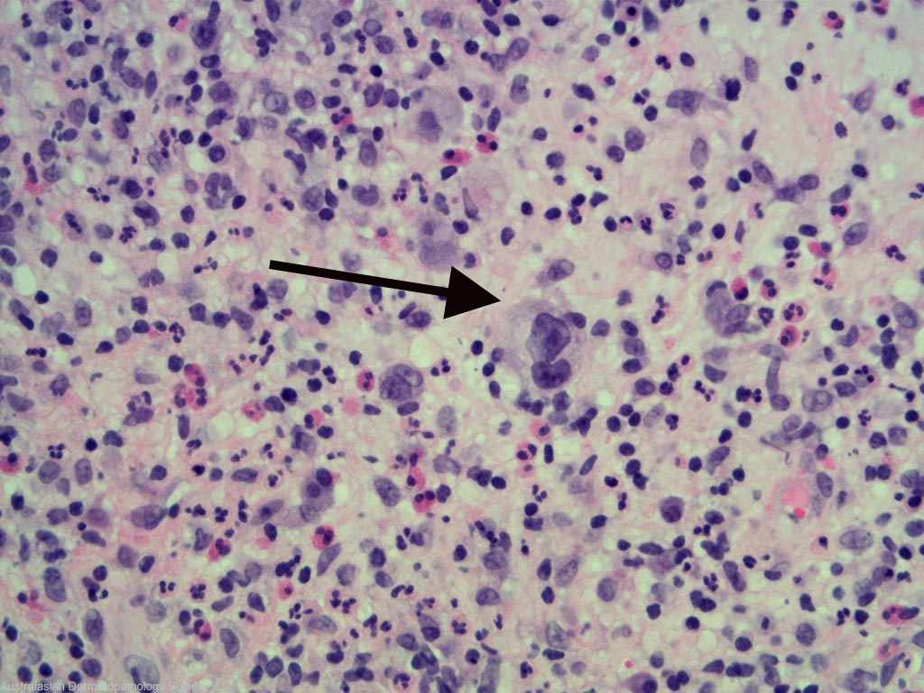
Description: Large atypical CD 30 +ve lymphocytes
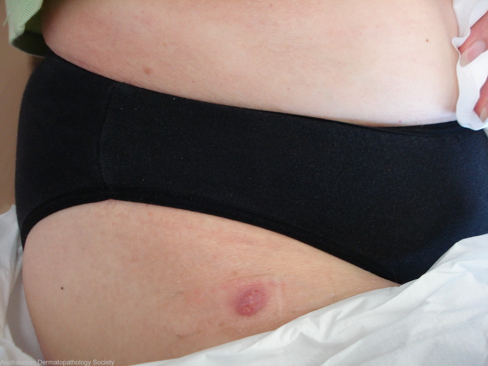
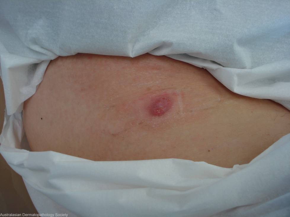
Clinical Features: Nodule,purple
Pathology/Site Features: Lymphohistiocytic infiltrate
Sex: F
Age: 62
Submitted By: Ian McColl
Differential DiagnosisHistory: 3465dmg This lady has had a skin rash clinically compatible with a T cell lymphoma but biopsies have only shown a chronic superficial dermatitis. Recently she has developed some nodules, 2 of which have spontaneously resolved but this one is increasing in size. Punch biopsy from the centre. DD Lymphomatoid papulosis, T cell lymphoma.
Description: Large atypical CD 30 +ve lymphocytes
Comments: Sections show a biopsy of skin in which there is a mixed infiltrate of C cells in the upper and mid dermis. The overlying epidermis is Excoriated and adjacent to this area shows some reactive hyperplasia. The infiltrate comprises lymphocytes, eosinophils and some neutrophils. Occasional histiocytes are also noted. Some plasma cells are also seen. The lymphocytes are mainly small in size although some medium sized lymphocytes and some larger quite atypical lymphocytes are present. These large atypical lymphocytes are positive for the CD30 immunoperoxidase stain. Most of the lymphocytes are positive for the CD3, CD5 and CD4 immunoperoxidase stains. The morphology and immunoprofile confirms a diagnosis of lymphomatoid papulosis (type A).


