
Diagnosis: Mycosis fungoides
Description: Diffuse infiltrated skin
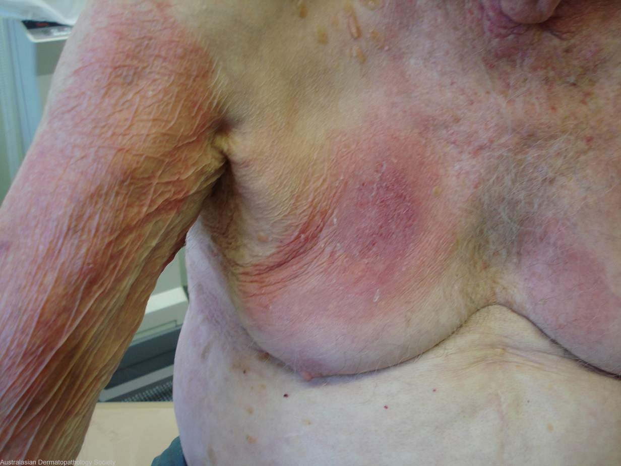
Clinical Features: Plaque
Pathology/Site Features: Arm,upper
Sex: M
Age: 82
Submitted By: Ian McColl
Differential Diagnosis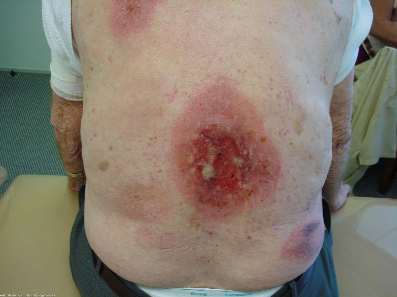
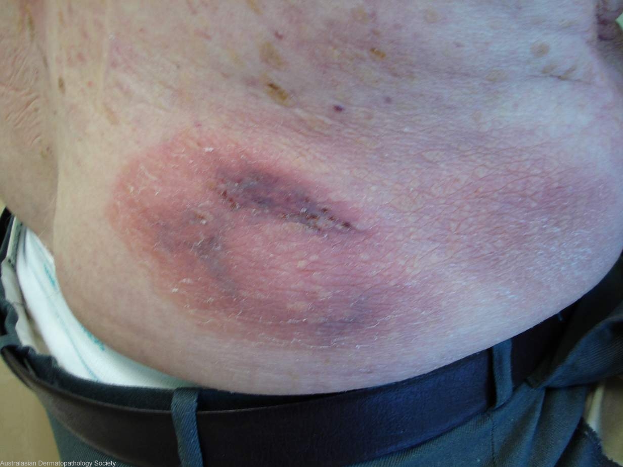

History: 82 years old male with these ulcerated plaques and patches scattered over various body areas. They arose over an 18 month period. Diagnosis Mycosis fungoides
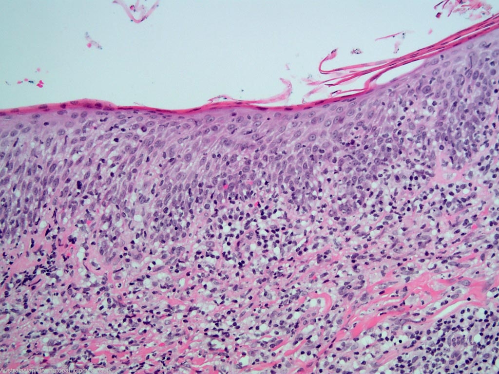
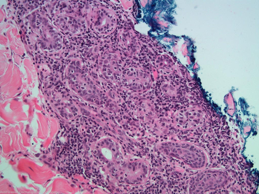
Description: Lymphocytes involving adnexae
Comments: Sections show a biopsy of skin in which there is a moderately dense infiltrate of mildly atypical lymphocytes involving the upper, mid and deep dermis. The lymphocytes show prominent epidermotropism as well as tight aggregation around pilosebaceous follicles and eccrine ducts. In addition to the lymphocytes occasional plasma cells and some small epithelioid granulomas are noted. The features present are those of plaque stage of mycosis fungoides.