
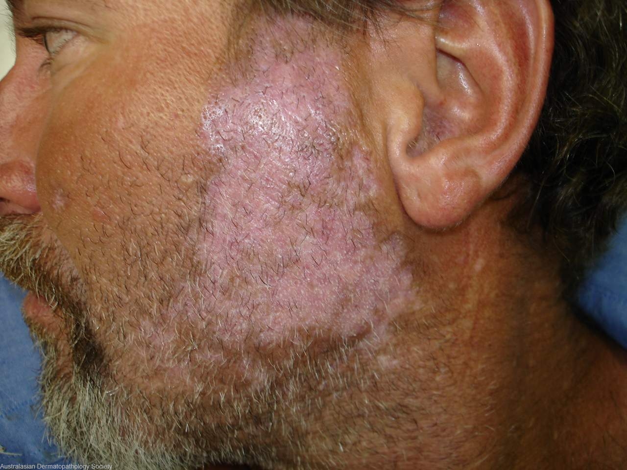
Diagnosis: Discoid lupus erythematosus
Description: Persistent red scaly plaque on cheek
Clinical Features: Plaque
Pathology/Site Features: Cheek
Sex: M
Age: 54
Submitted By: Ian McColl
Differential Diagnosis

History: 5513ca Male with a 2 year history of a red scaly plaque on both cheeks with hair loss in the beard. Treated by GP as fungal infection. Clinically discoid lupus erythematosus. May also have porphyria cutanea tarda. Two biopsies
Description: Discoid lupus erythematosus
Comments: Sections show a biopsy of skin in which there is a quite mild lichenoid reaction involving the surface epithelium and a more pronounced lichenoid reaction involving follicular epithelium. There is increased thickness of the epidermal basement membrane. The upper dermis shows moderate solar elastosis. There is an upper, mid and deep dermal perivascular and perifollicular infiltrate of lymphocytes, histiocytes and some plasma cells. Some follicular plugging is also noted. The features present are those of discoid lupus erythematosus.