
Diagnosis: Desmoplastic Melanoma
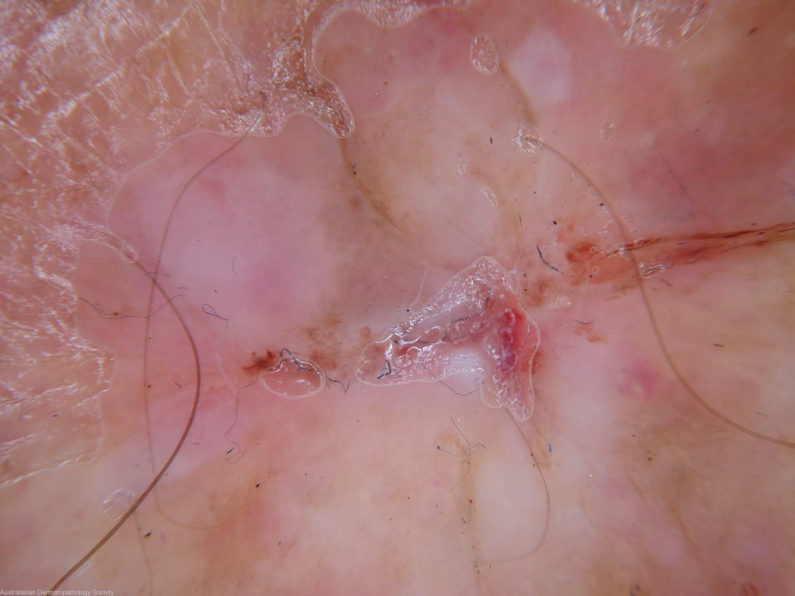
Description: Really little to see
Clinical Features: Nodule pink
Pathology/Site Features: Dermatoscopy
Sex: M
Age: 65
Submitted By: Ian McColl
Differential Diagnosis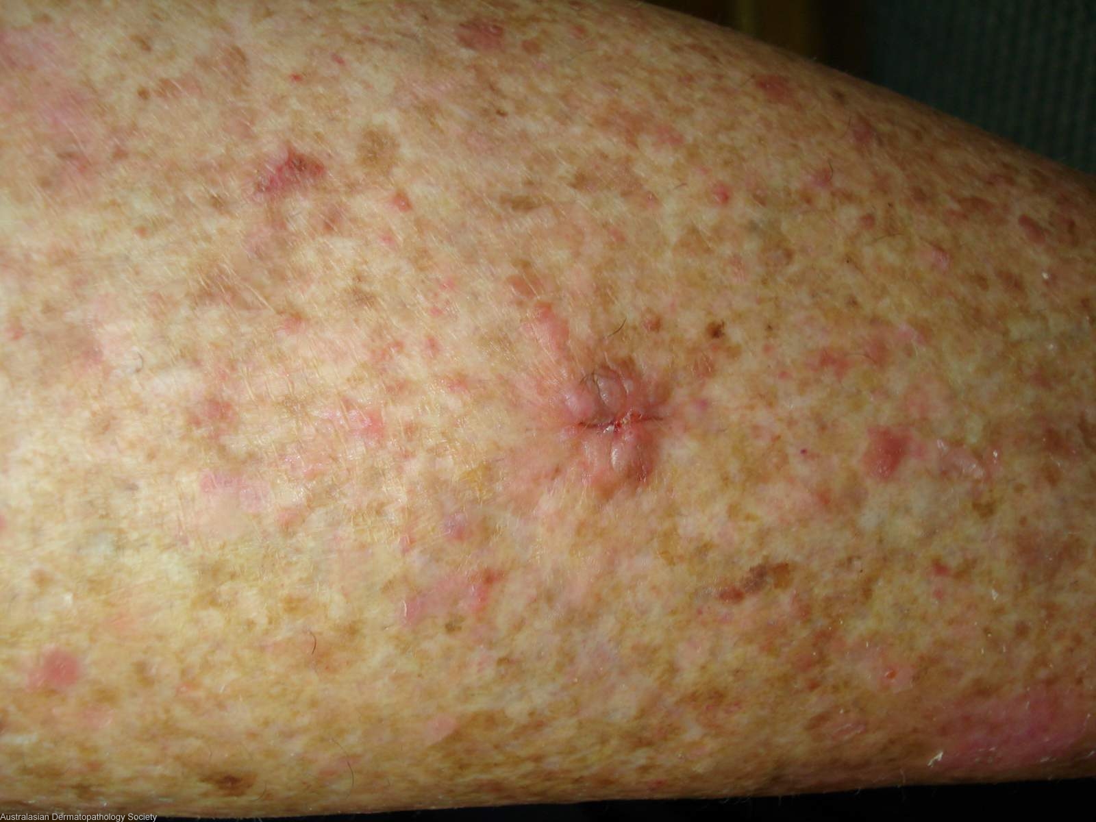

History: 0786ra 4 month history of a lesion on the shin. Clinically I thought it was a BCC but queried an amelanotic melanoma as the differential diagnosis.
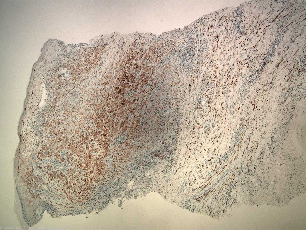
Description: S100 stain demonstrating deeply invasive melanocytes
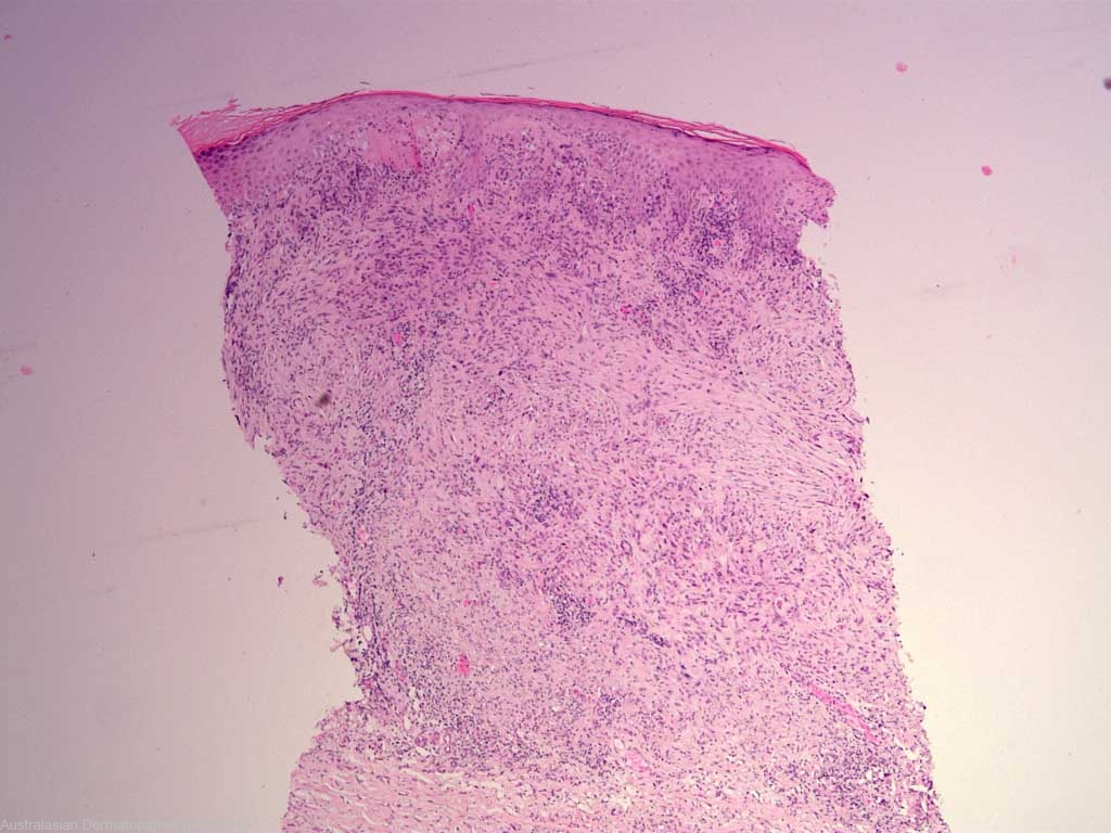
Comments: Sections show a biopsy of skin in which there is a proliferation of atypical spindle cells occupying most of the dermis. These have irregular nuclei and mitoses are easily found. They are accompanied by a patchy infiltrate of lymphocytes. In the overlying epidermis, there appear to be atypical melanocytes in the basal epidermis. The overall features favour a desmoplastic malignant melanoma. It is level 4 and 4.0 mm in greatest depth.