
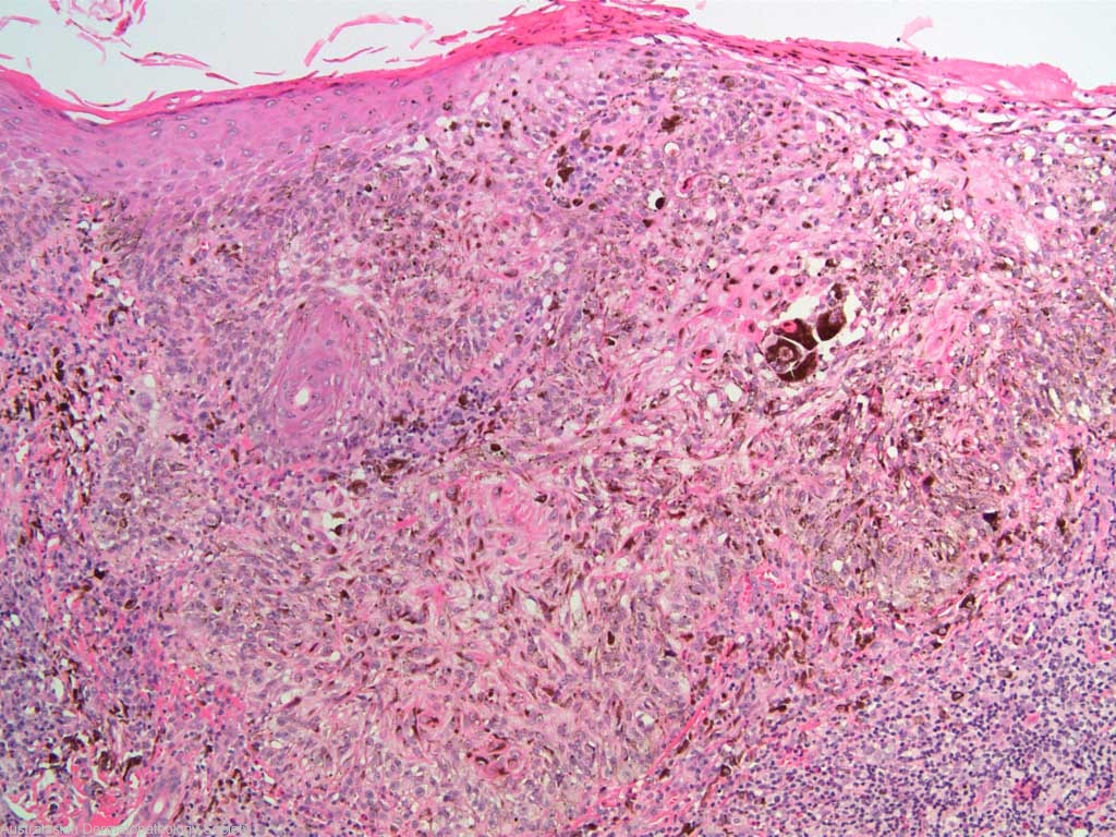
Diagnosis: Melanoma
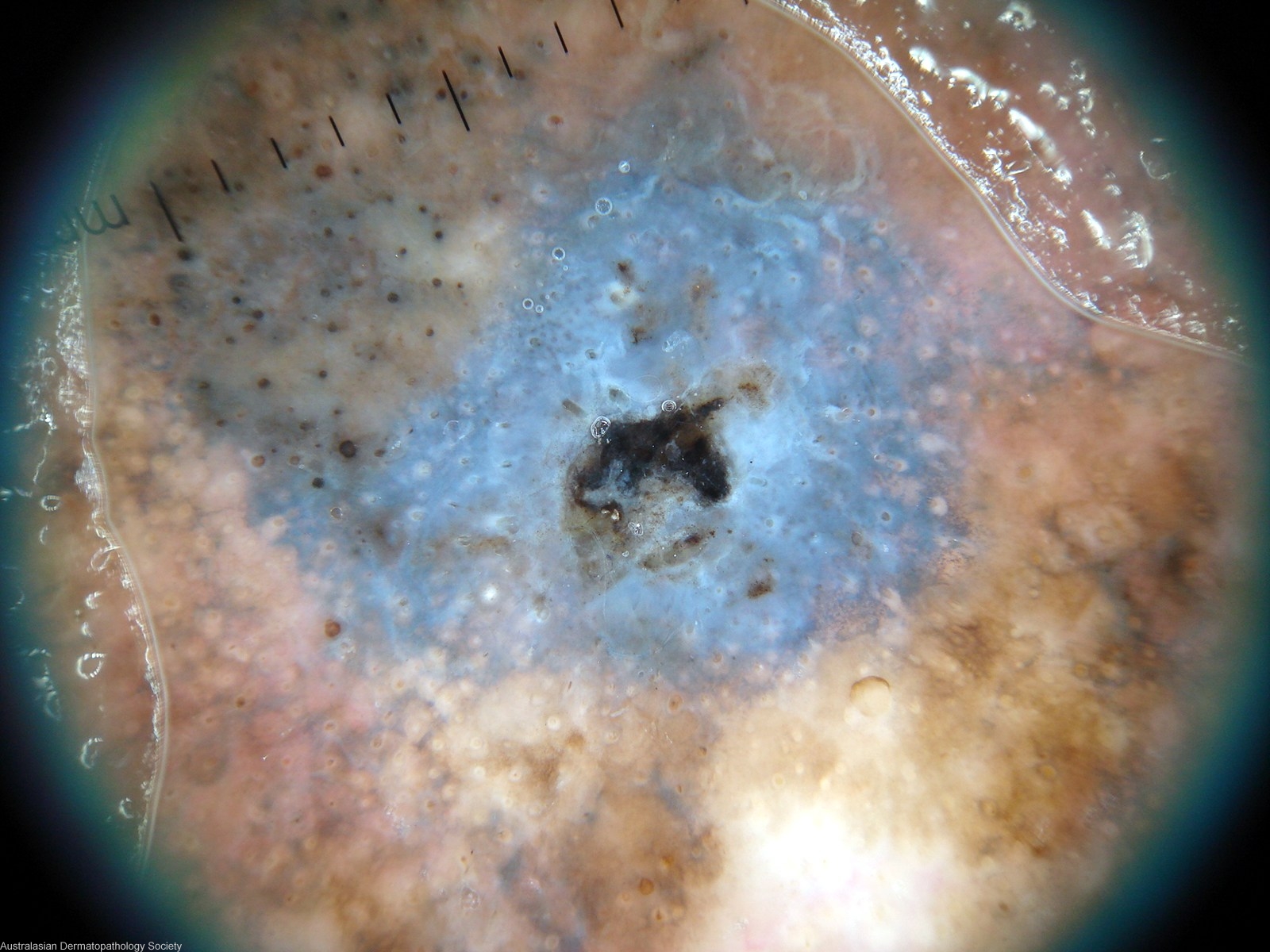
Description: Blue colour represents dermal invasion
Clinical Features: Macule black
Pathology/Site Features: Dermatoscopy
Sex: M
Age: 68
Submitted By: Ian McColl
Differential Diagnosis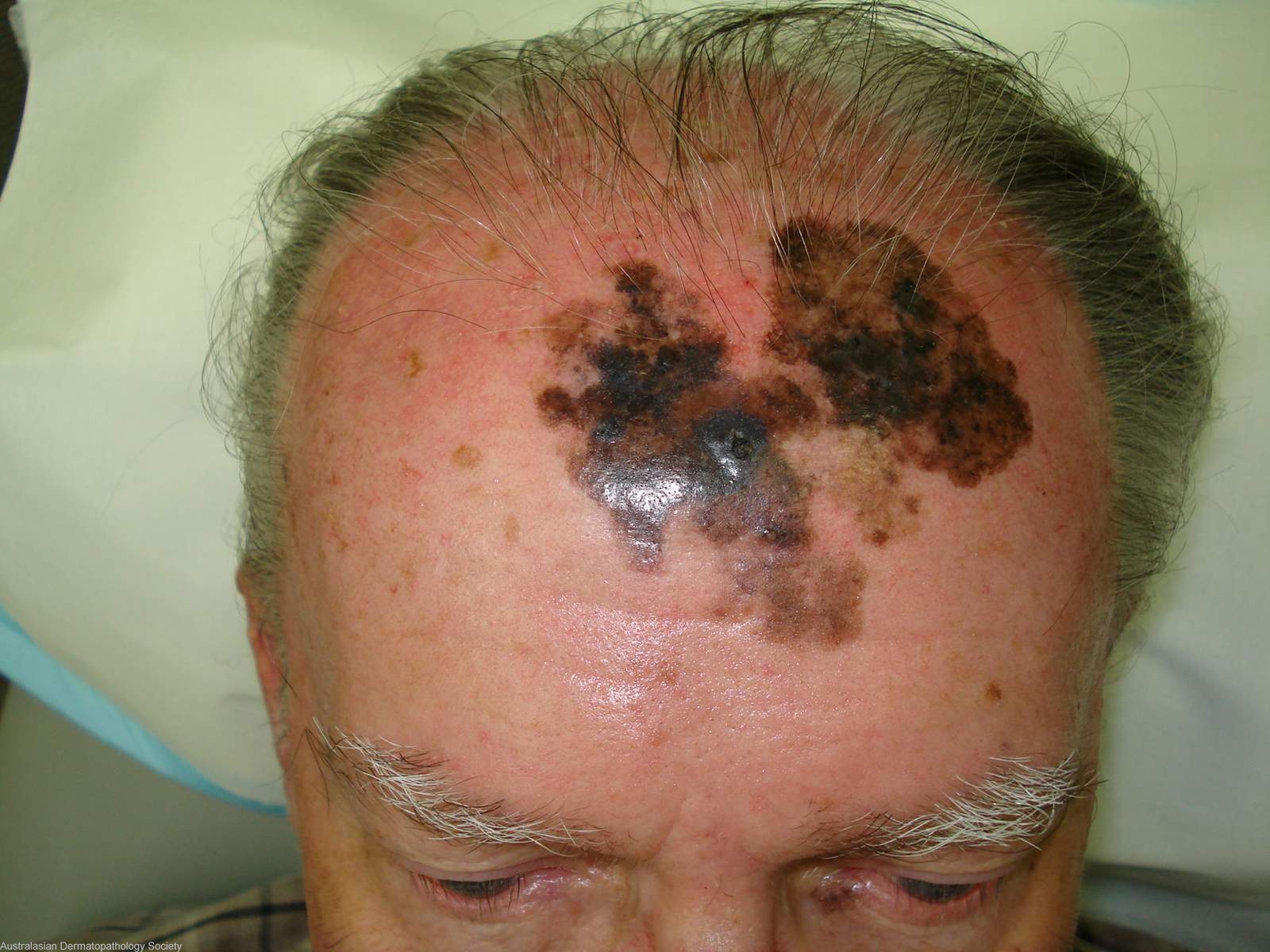
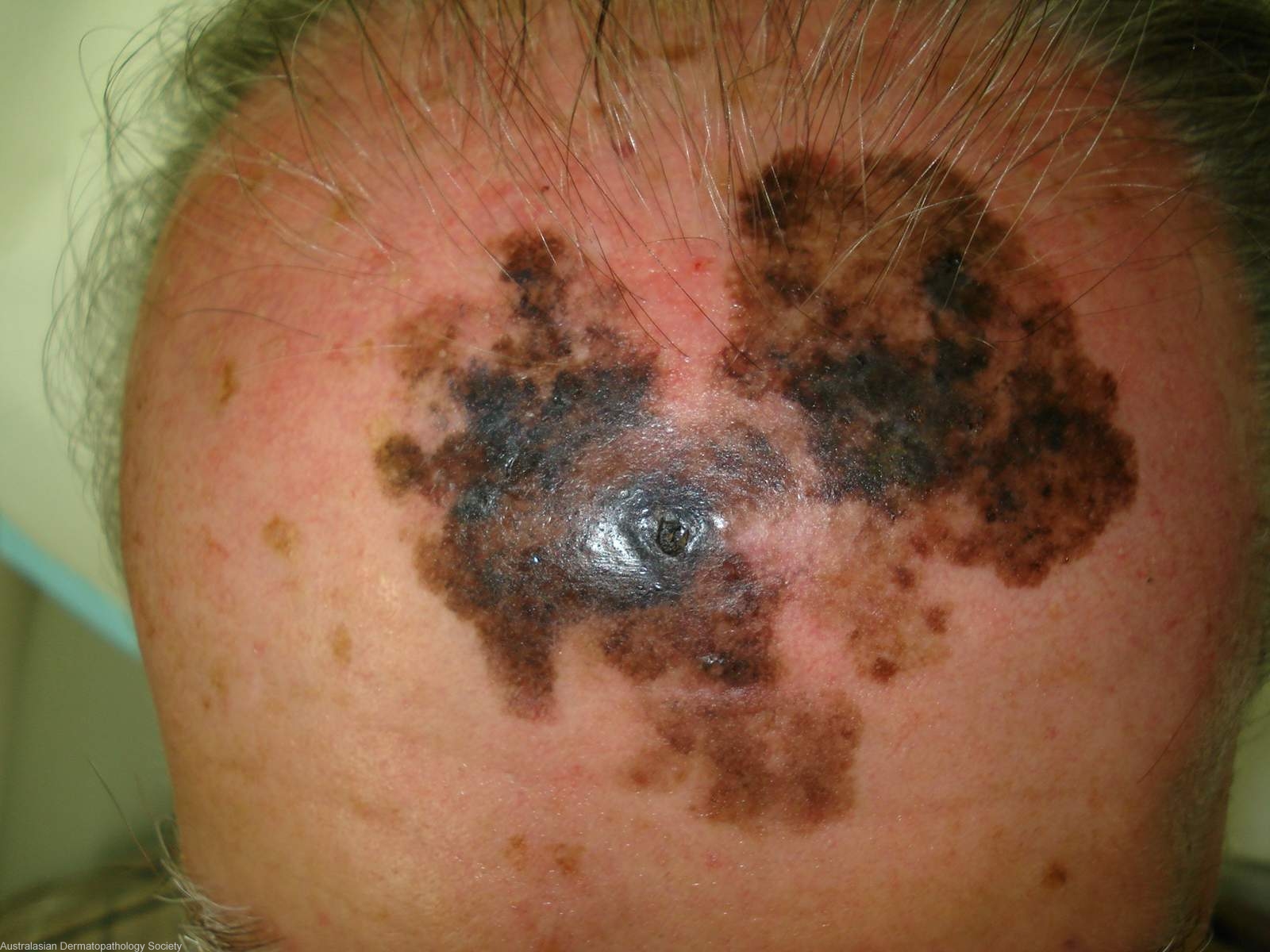

History: 5654aw This lesion has been slowly growing over a period of 16 years. Never previously biopsied or treated. Clinically lentigo maligna melanoma. Biopsies taken from 3 areas across the lesion. The dermatoscope image is from the central area showing the blue colour of deep dermal melanin.
Description: Lentigo maligna
Comments: Invasive malignant melanoma is present in all 3 biopsies. The presence of some lentigo maligna at the edge of specimen 3 allows the designation of lentigo maligna melanoma. The thickness of the invasive component in the three biopsies varies between 0.6mm and 1.2 mms.