
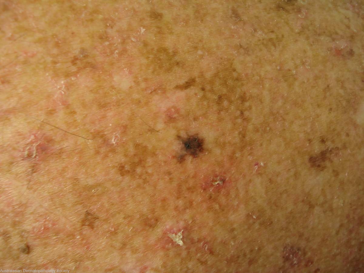
Diagnosis: Melanoma
Description: Black macule on the back
Clinical Features: Macule black
Pathology/Site Features: Back
Sex: M
Age: 82
Submitted By: Ian McColl
Differential Diagnosis

History: 1160kk Atypical pigmented lesion on the upper back suggestive of melanoma. Dermatoscopy image included. Lab reference 567671110
Description: malignant melanoma in-situ
Comments: The biopsy shows a malignant melanoma in-situ. An atypical nested proliferation of melanocytes shows full thickness spread through the epidermis. There is surface hyperkeratosis. The dermis shows fibroplasia and scattered inflammatory infiltrate with pigmented laden macrophages suggesting regression. There is no unequivocal dermal component to the lesion.