
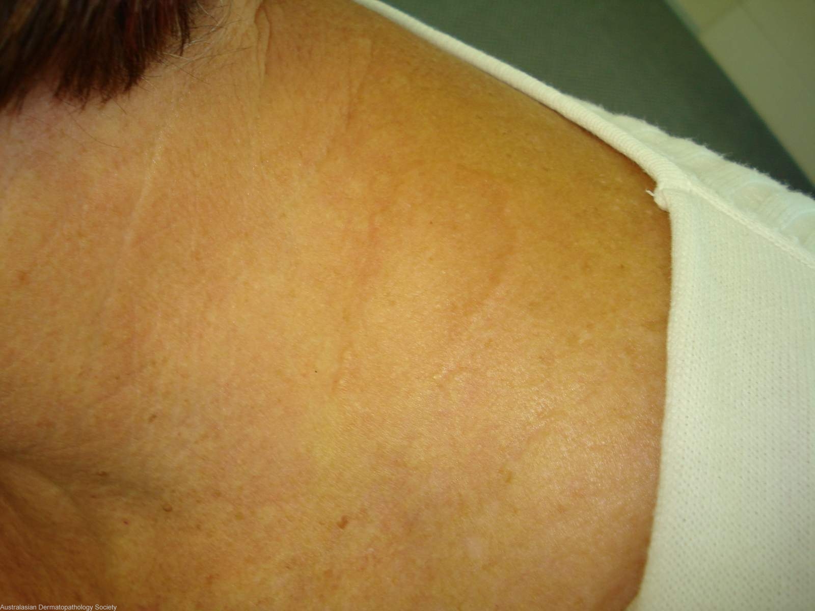
Diagnosis: Actinic granuloma
Description: Annular plaque on side of neck
Clinical Features: Annular
Pathology/Site Features: Neck side
Sex: F
Age: 65
Submitted By: Ian McColl
Differential Diagnosis


History: 4015at Annular plaque on shoulder and neck for 12 months. No response to topical and oral antifungals. DD Actinic granuloma Biopsies from middle and edge of lesion.
Description: central biopsy (elastic stain) - loss of elastic fibres
Comments: Sections from the edge biopsy show a granulomatous reaction involving the upper and mid dermis. The granulomas consist of both mononucleated and multinucleated histiocytes of foreign body giant cell type. Fragments of elastotic fibres are present within the granulomas. Sections from the central biopsy show no granulomas. There is however loss of elastotic material and elastic fibres from the upper dermis. The changes present are those of actinic granuloma. Loss of elastic fibres from the central part of the lesion is typical of this condition.