

Diagnosis: Melanoma
Description: Pigmented lesion on leg varying in colour
Clinical Features: Macule black
Pathology/Site Features: Leg
Sex: M
Age: 77
Submitted By: Ian McColl
Differential Diagnosis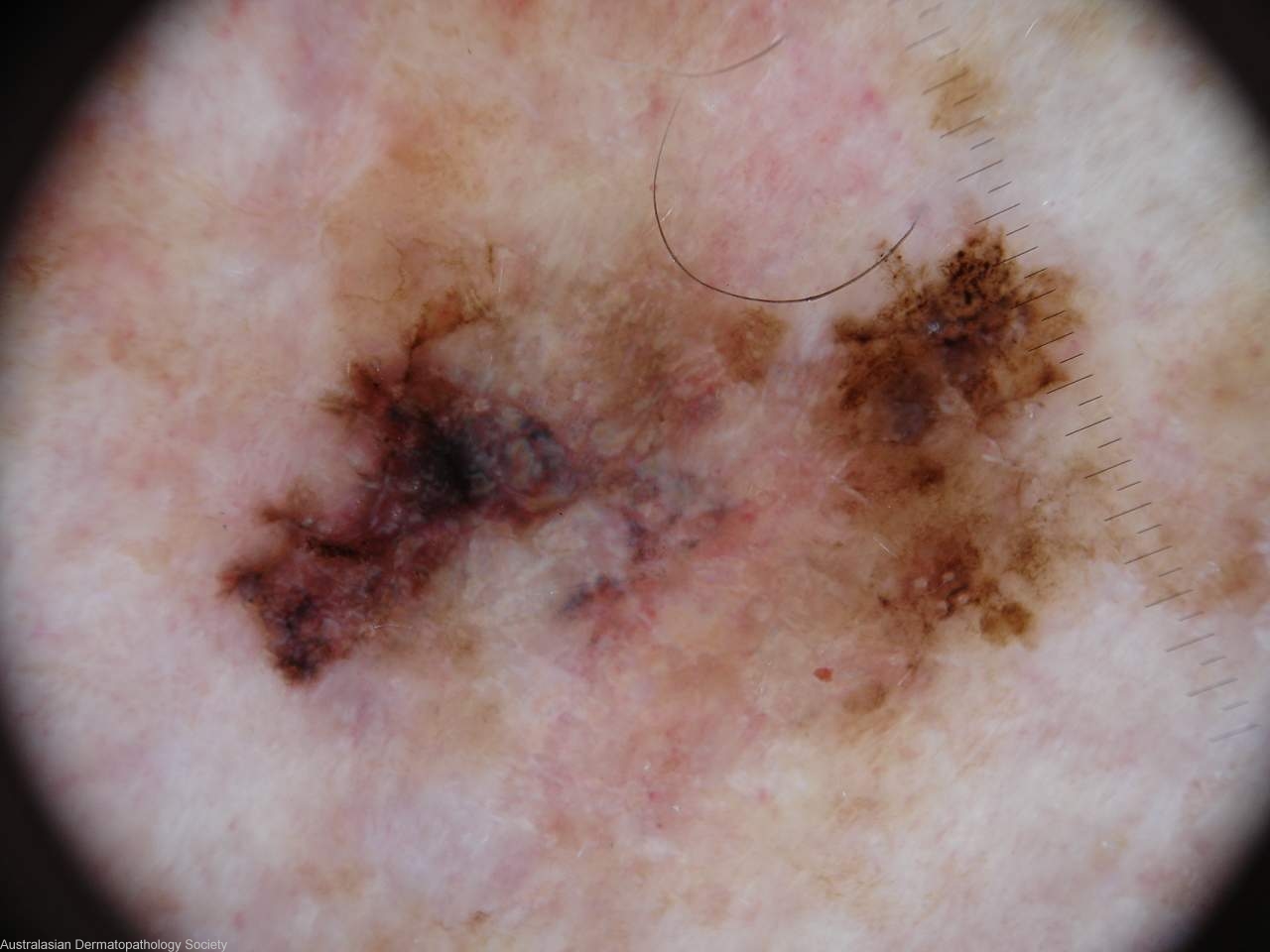
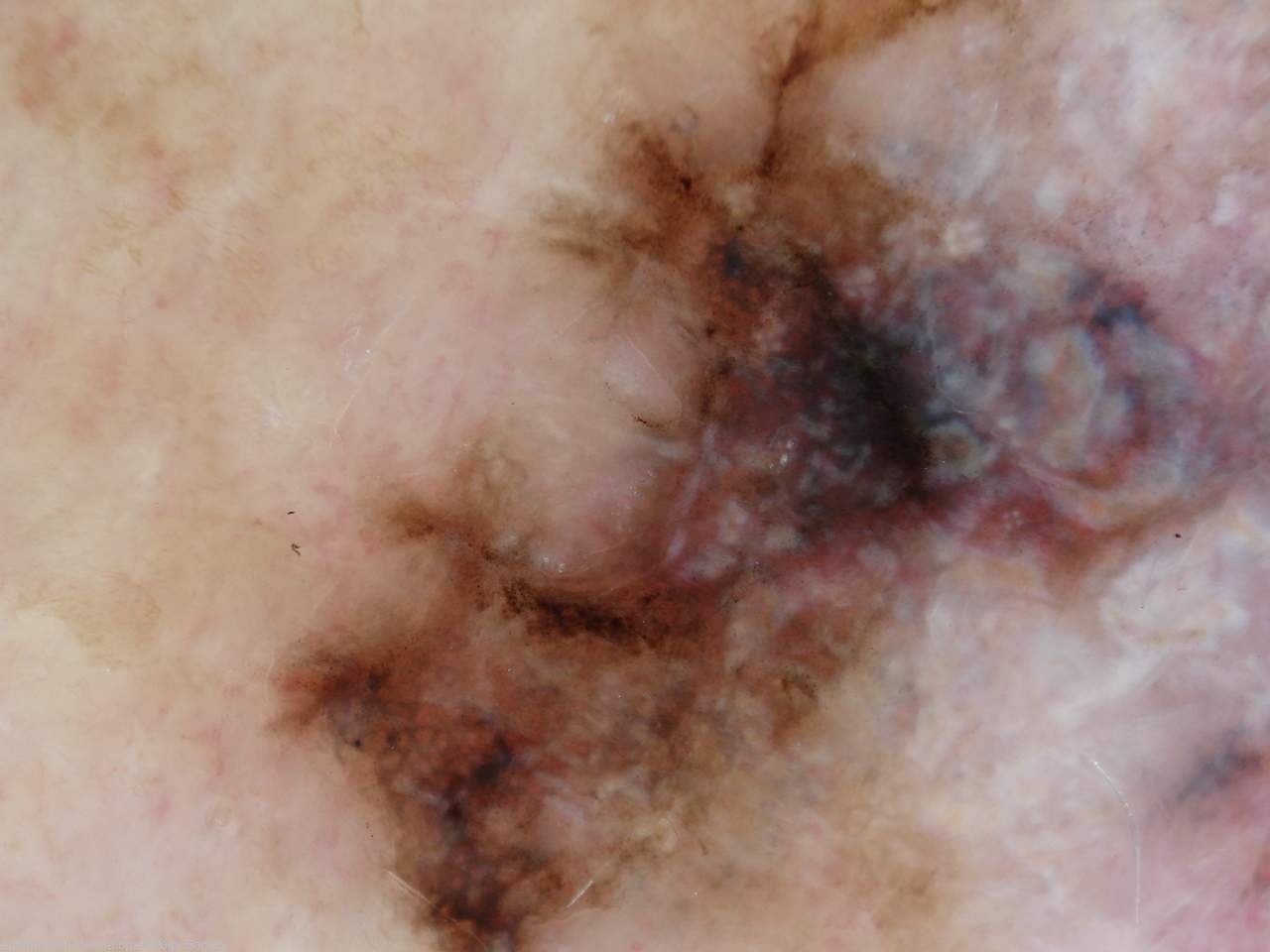
History: 5557lk 77 years old male with this pigmented lesion on his leg varying in size and colour over two years. Clinically a regressed superficial spreading melanoma. Incisional biopsy taken of the dark left edge.
Description: Regressed area
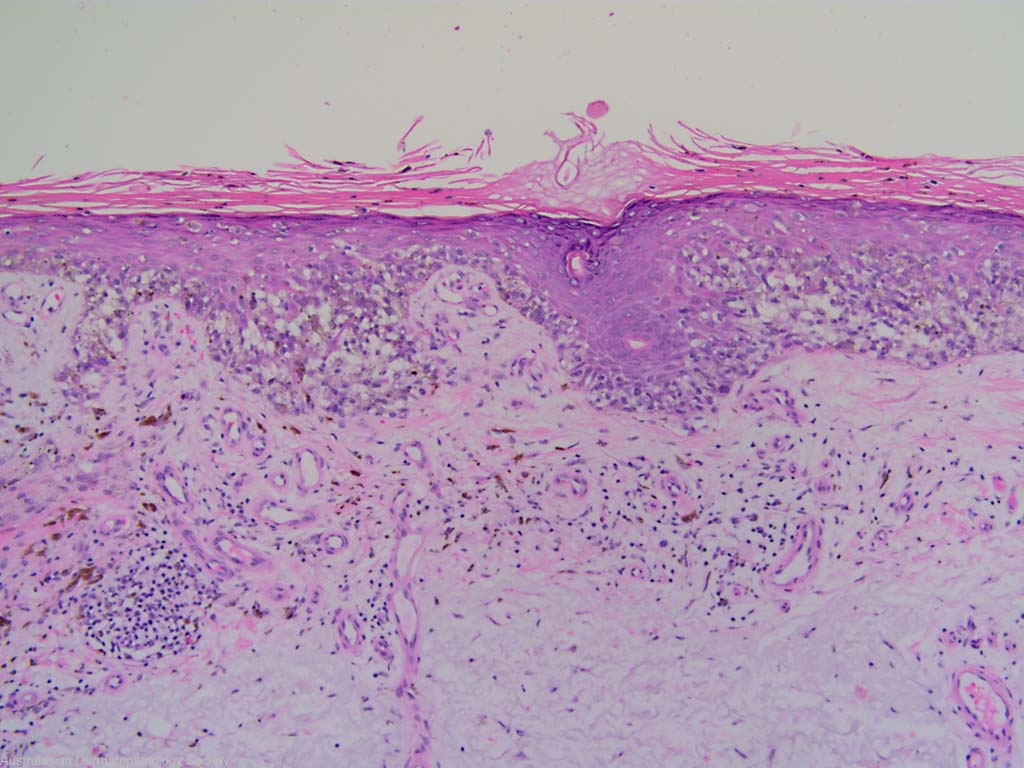
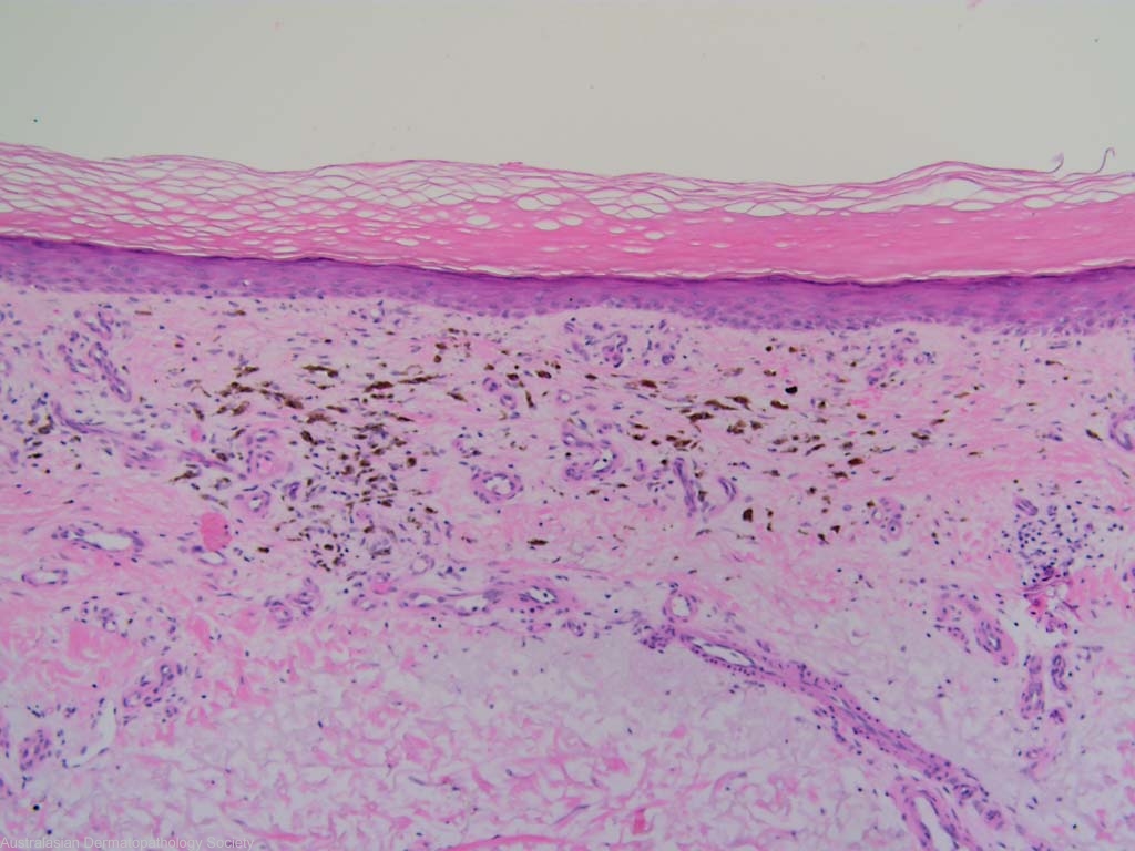
Comments: Sections show a biopsy of a superficial spreading malignant melanoma. It is predominantly Level 1 ( in situ) although there is a focus of dermal invasion (Level 2; 0.40mm in greatest depth). There is evidence of old regression with epidermal atrophy, loss of melanocytes, upper dermal fibrosis and upper dermal pigment incontinence.