
Diagnosis: Dysplastic lentiginous nevus
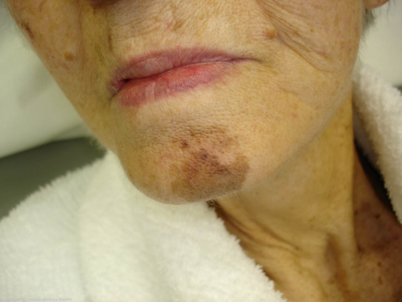
Description: Dark slowly growing macule on the chin
Clinical Features: Macule black
Pathology/Site Features: Chin
Sex: F
Age: 82
Submitted By: Ian McColl
Differential Diagnosis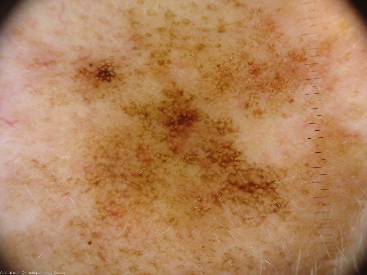
History: 2429mm Lady in her 80s with this pigmented lesion on her chin. Dermatoscope image shown. Biopsy taken from the darkest area superiorly and from the more uniform area below. DD HMF or melanoma in situ
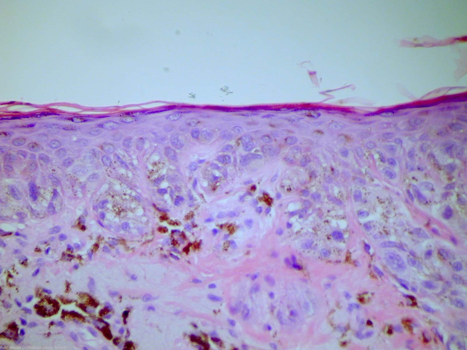
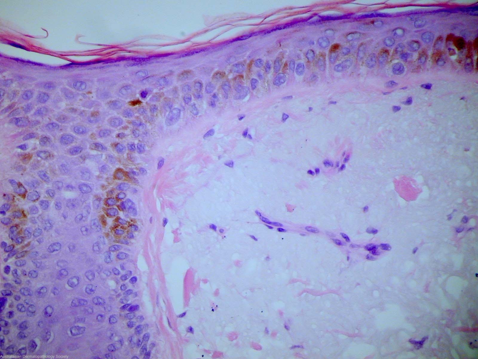
Description: lentiginous proliferation
Comments: Sections show a biopsy of skin in which there is a proliferation of atypical melanocytes restricted to the basal epidermis. They form both a lentiginous as well as nested pattern. There is no evidence of upward epidermal spread and no evidence of dermal involvement. The dermis shows marked solar elastosis. The features best fit for a dysplastic junctional lentiginous naevus. There is no evidence of malignancy.
Editor - With PRAME and other studies we now call these lentiginous melanomas.