
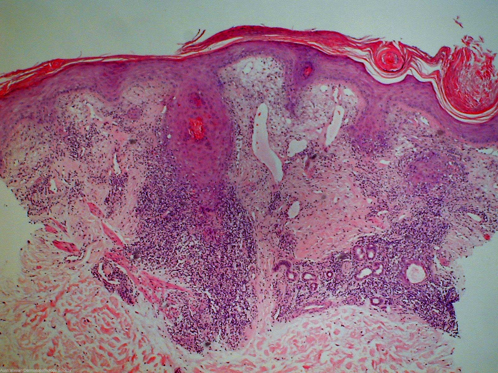
Diagnosis: Discoid lupus erythematosus
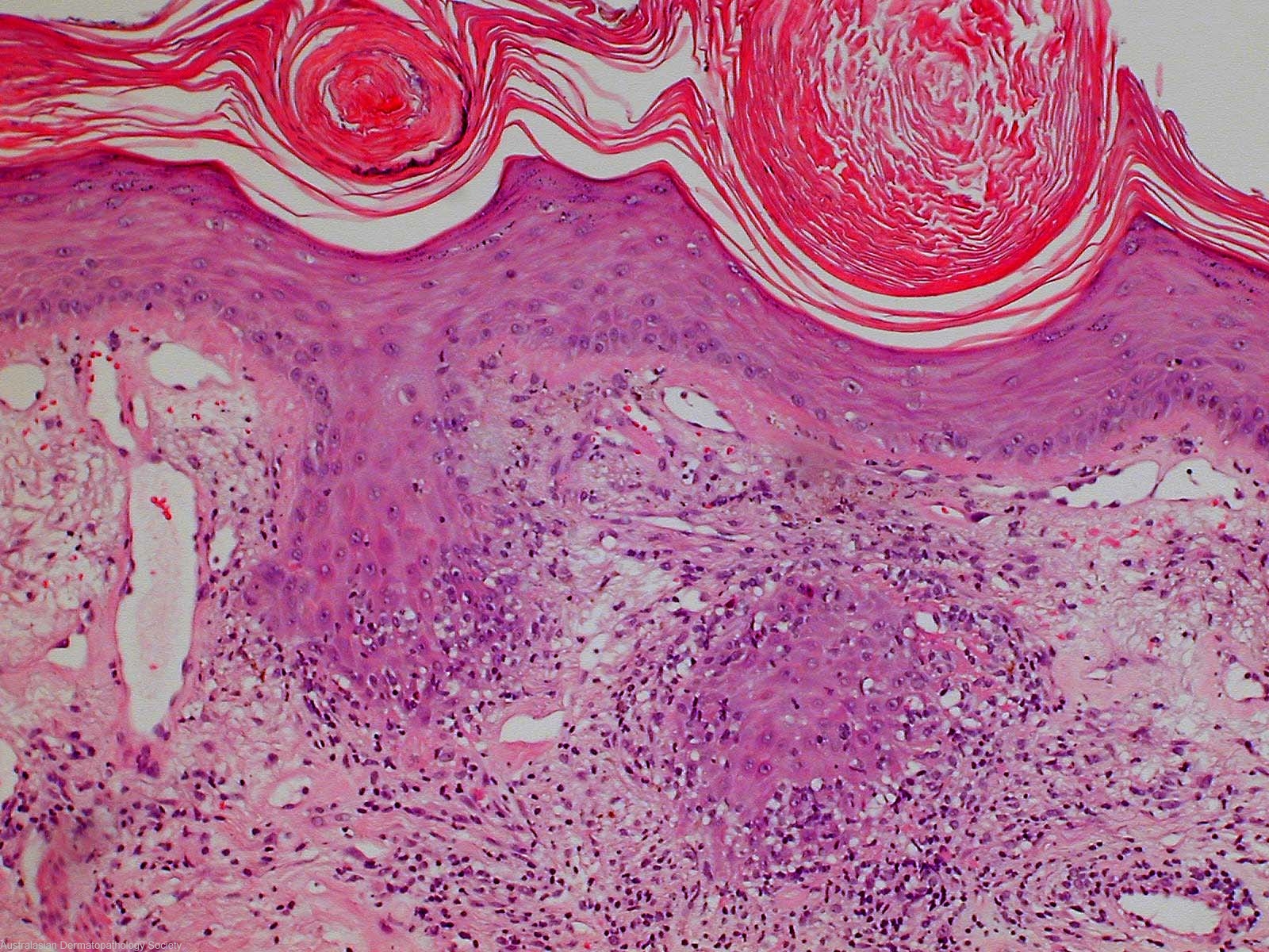
Description: Biopsies of skin in which there is a lichenoid reaction involving the epidermis and follicular epithelium. This is accompanied by a superficial and mid dermal perivascular and perifollicular infiltrate of lymphocytes
Clinical Features: Red,scaly
Pathology/Site Features: Periadnexal infiltrate
Sex: F
Age: 30
Submitted By: Ian McColl
Differential Diagnosis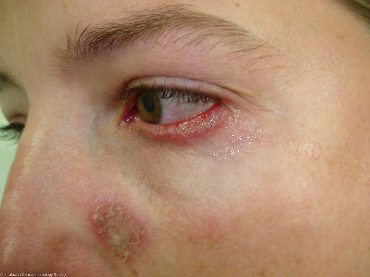
History: 4826kh Two year history of indurated areas on both lower eyelids followed by plaques x 2 on both cheeks. No steroid creams used as far as I can ascertain. DD Discoid lupus, sarcoidosis or necrobiotic xanthogranuloma. Biopsies cheek plaque and lower eyelid
Description: Folliculo-centric lichenoid reaction
Comments: Sections show a biopsies of skin in which there is a lichenoid reaction involving the epidermis and follicular epithelium. This is accompanied by a superficial and mid dermal perivascular and perifollicular infiltrate of lymphocytes. There is thickening of the epidermal basement membrane and some overlying ortho- and parakeratosis. The changes are those of discoid lupus erythematosus.