

Diagnosis: Subacute lupus erythematosus
Description: Red non scaly annular rash back
Clinical Features: Annular
Pathology/Site Features: Back
Sex: F
Age: 78
Submitted By: Ian McColl
Differential Diagnosis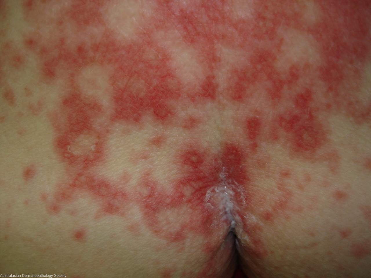
History: 2239mr Recent case previously thought to be a drug reaction with impetiginisation but when dose of steroids reduced this rash came up. Previous immunofluorescence negative. DD Lupus erythematosus, still drug reaction, drug induced lupus?
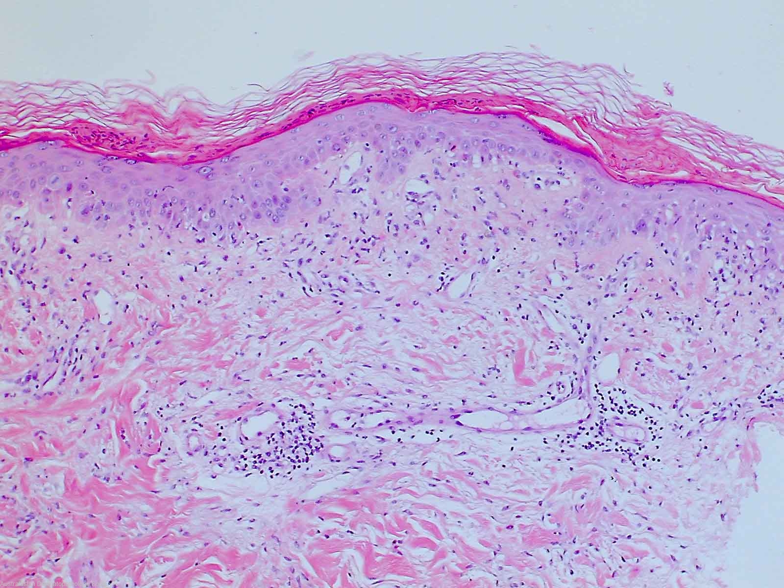
Description: Lichenoid dermatitis
Comments: Sections show biopsies of skin in which there is a lichenoid reaction present. There is thinning of the epidermis with basal epidermal vacuolar change and quite prominant Civatte bodies. There is overlying patchy parakeratosis. There is a mild to moderate superficial and mid dermal perivascular infiltrate of lymphocytes. These features best fit for a subacute type of lupus erythematosus.