
Diagnosis: Dysplastic lentiginous nevus
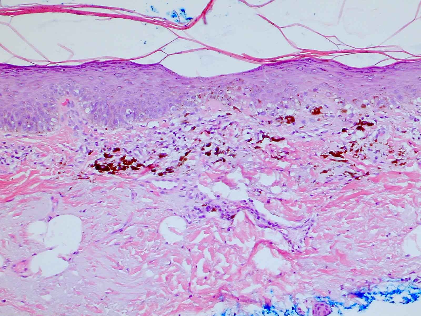
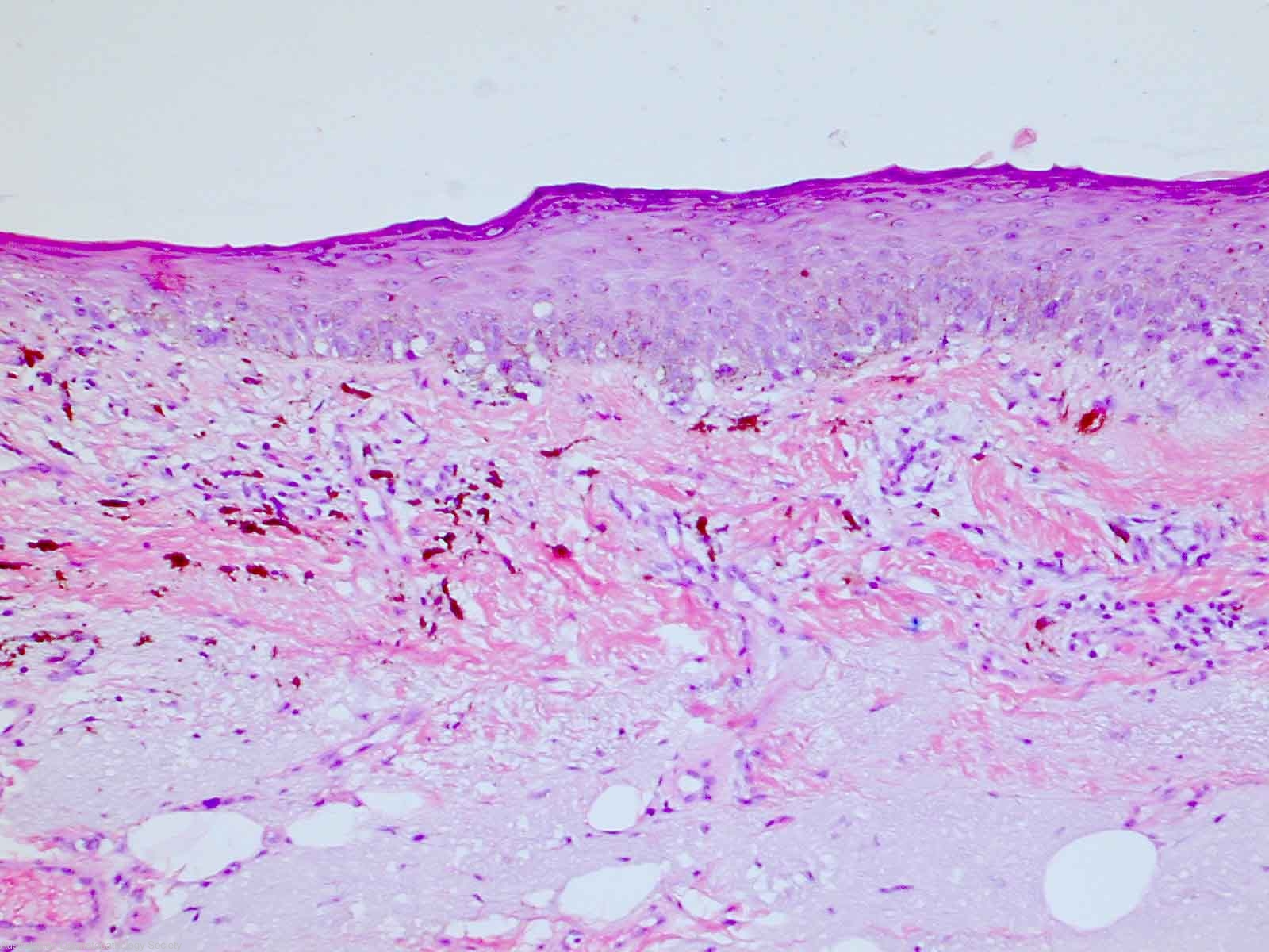
Description: There is extensive upper dermal fibrosis and pigment incontinence indicative of some degree of previous regression.
Clinical Features: Macule brown
Pathology/Site Features: Pigment incontinence
Sex: M
Age: 80
Submitted By: Ian McColl
Differential Diagnosis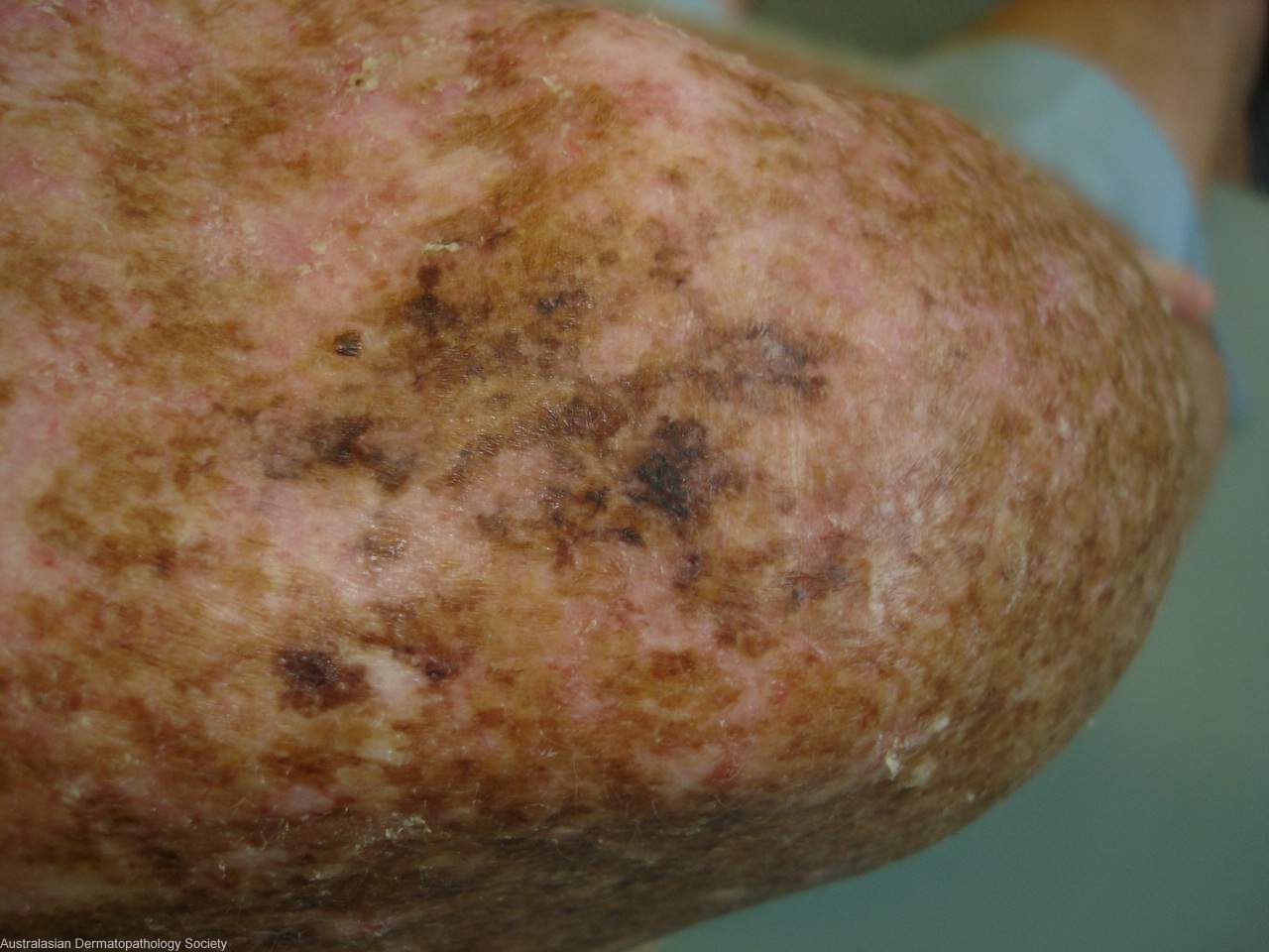
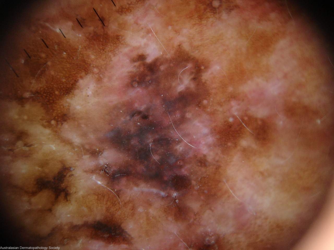
History: 4619th Severly sun damaged back in an elderly male. Shave biopsy of area shown in dermoscopy image.
Description: Pigmented area right shoulder
Comments: Sections show a lesion consisting of a proliferation of atypical melanocytes in the basal epidermis. They form a predominantly lentiginous pattern with only a few nests. There is extensive upper dermal fibrosis and pigment incontinence indicative of some degree of previous regression. There is no evidence of upward epidermal spread and no dermal involvement by the atypical melanocytes. I think it best to regard the lesion as a type of dysplastic junctional lentiginous naevus with regression.