
Diagnosis: Xanthogranuloma
Description: Dense dermal infiltrate of foamy histiocytes with some lymphocytes and eosinophils
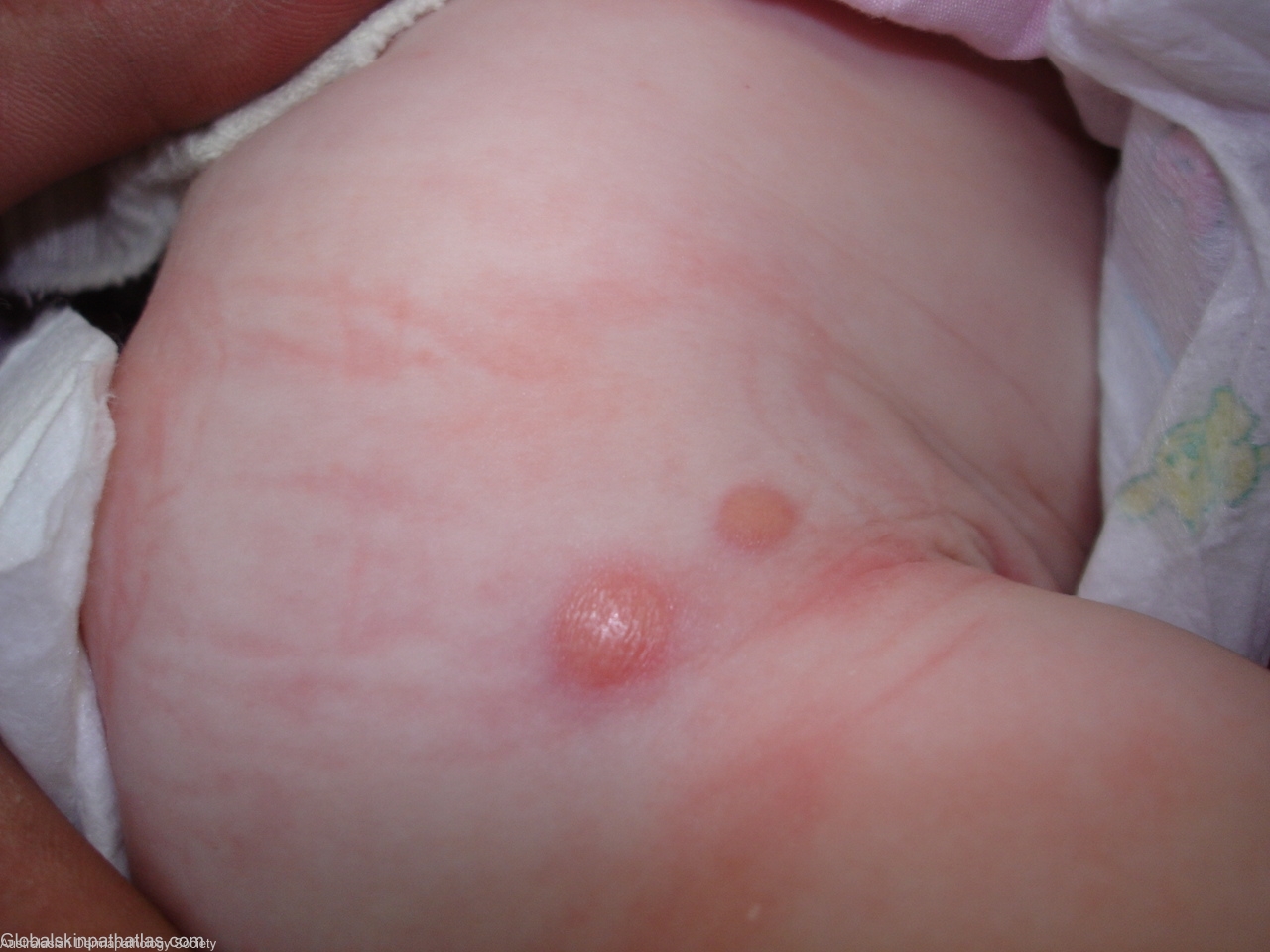
Clinical Features: Nodule
Pathology/Site Features: Foam cells
Sex: M
Age: 0.3
Submitted By: Ian McColl
Differential Diagnosis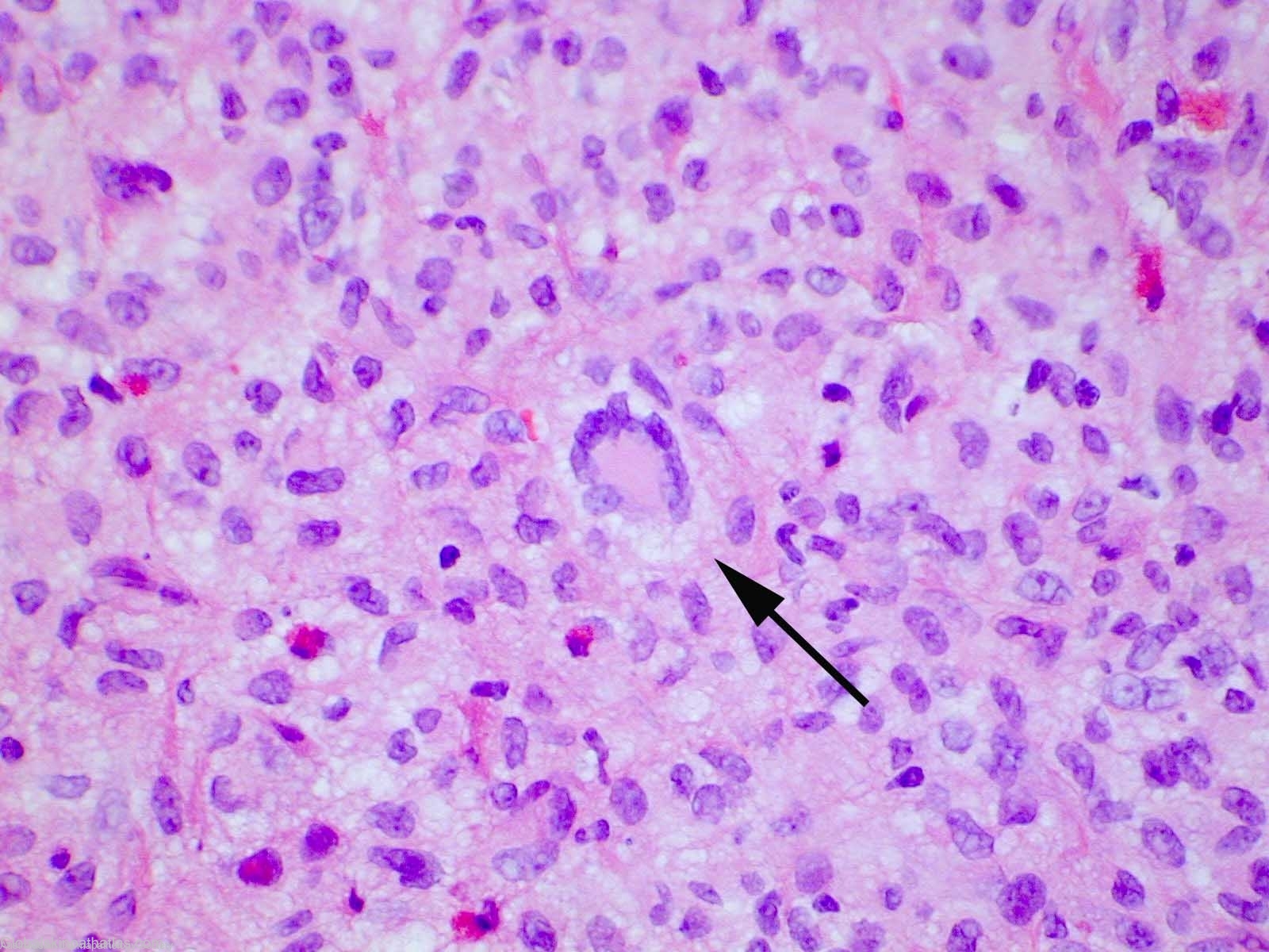
History:
4 month old child who was born with the lower nodule.It has doubled in size and the upper lesion has recently formed.No blistering on rubbing.DD Xanthogranuloma,Mastocytoma.Punch biopsy through centre of lower nodule.
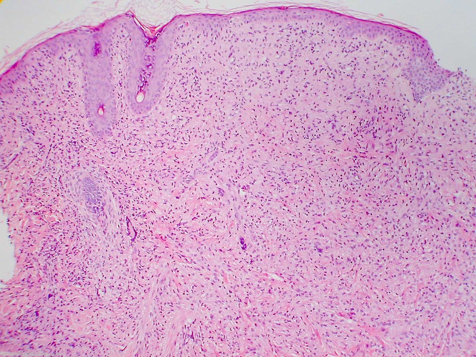
Pathology Description There is a dense infiltrate of histiocytes completely replacing the dermis. These are plump and vary from spindle shaped to round. Some of these histiocytes have foamy cytoplasm. Admixed with histiocytes are quite frequent Touton giant cells. In addition a number of lymphocytes and eosinophils are admixed with the histiocytes. The features present are those of a juvenile xanthogranuloma.