

Diagnosis: Blastic NK cell lymphoma
Description: Multiple black nodules on the lower leg
Clinical Features: Nodule,black
Pathology/Site Features: Leg
Sex: M
Age: 87
Submitted By: Ian McColl
Differential Diagnosis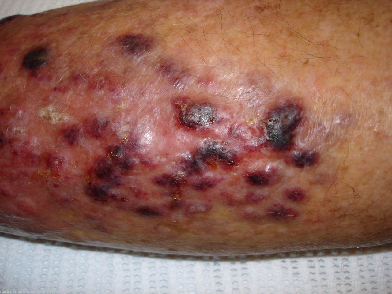
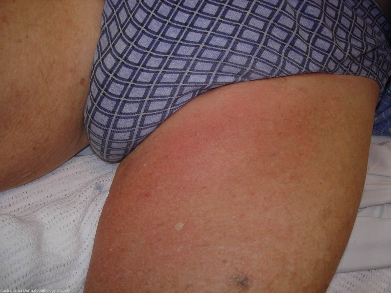
History: 0340hn 87 years old male with a 3 week history of a lesion on his shin diagnosed by a dermatologist as a keratoacanthoma who woke up one morning last week with this plaque on his leg with these multiple vascular like nodules. These have grown further in the last week with associated matted lymphadenopathy in the groin. He is not systemically unwell,mild fever,no lesions elsewhere, no known immunosuppression. Previous malignancies of lung,prostate and gut! DD Bacillary angiomatosis, Kaposi's sarcoma, Angiosarcoma, Sweets syndrome, Leukemia cutis, Metastatic melanoma. Tissue for histology and culture plus Warthin Starry stain
Description: Matted tender lymphadenopathy in the groin
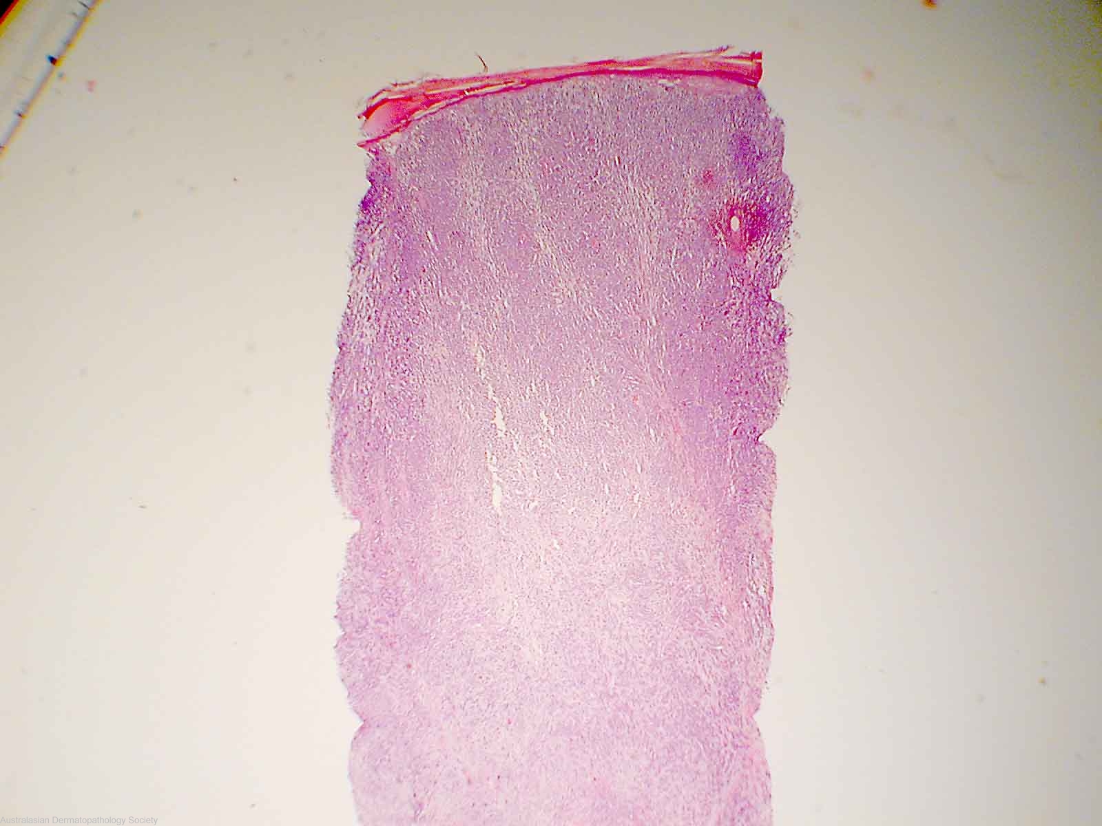
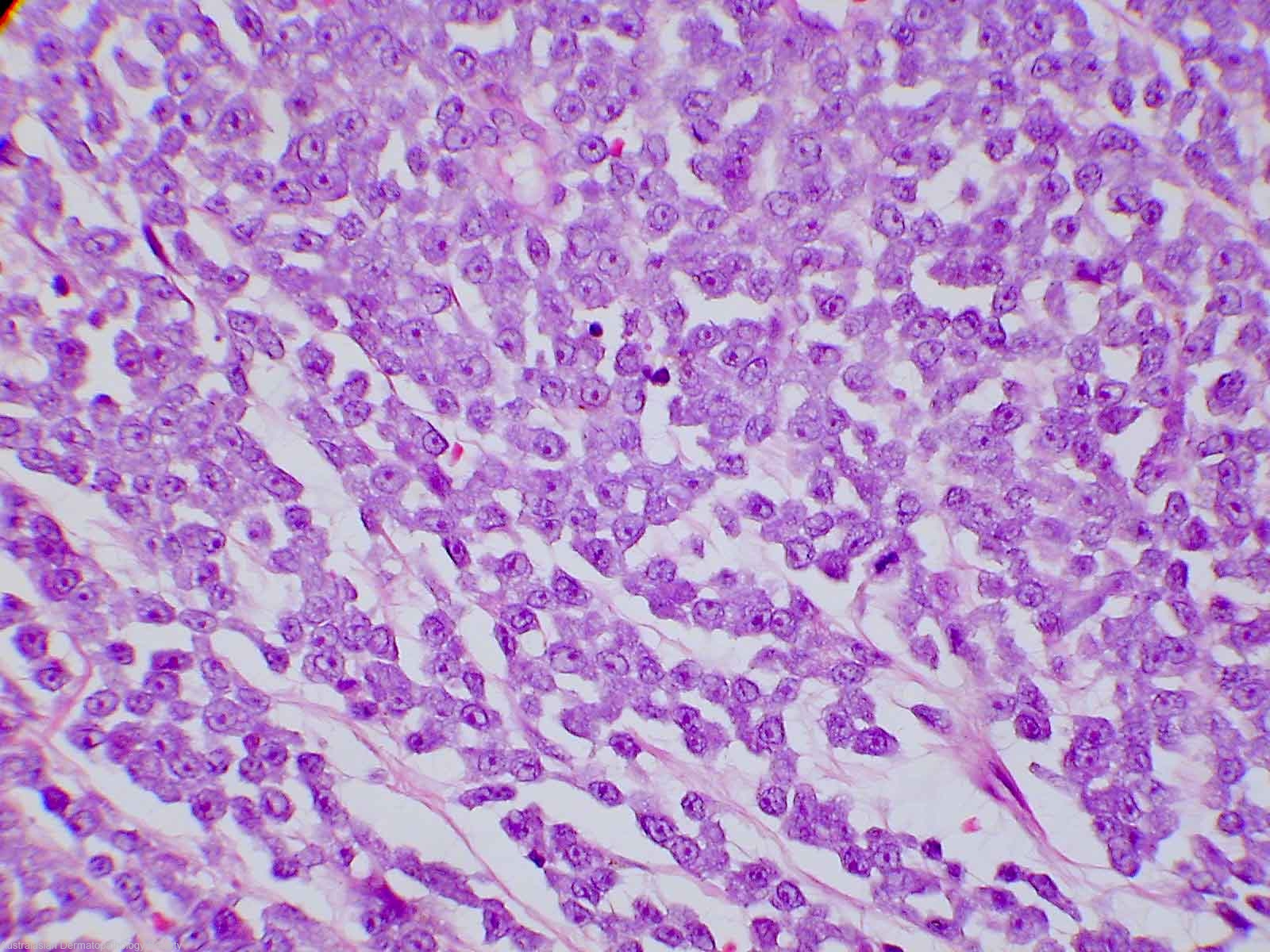
Comments: The dermis is completely replaced and infiltrated by malignant tumour cells. The cells are medium in size and have small amounts of grey cytoplasm. The cell nuclei are moderately pleomorphic and contain fairly prominent amphophilic nucleoli. The cells completely fill the dermis forming sheets with some cell discohesion. The mitotic rate is high. In addition tumour cells invade the overlying epidermis in a focally pagetoid fashion. The tumour cells are positive for the pan leucocyte marker CD45. They are also positive for the CD43 and CD56 immunoperoxidase stains. The cells are negative for the CD3, CD5, CD10, CD20, CD30, CD38, CK20, myeloperoxidase, TTF-1, HMB45, S100 and AE1/AE3 immunoperoxidase stains. The morphology and immuno-profile of the tumour indicates that it is a blastic non-Hodgkins lymphoma. The positive CD56 and CD43 staining with concomitant negative CD3 and myeloperoxidase staining indicates a most likely diagnosis of blastic NK-cell lymphoma.
View Full Size