
Diagnosis: Mycosis fungoides
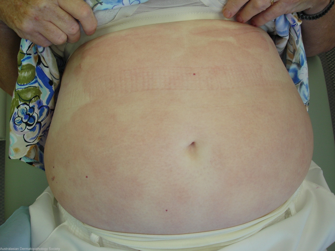
Description: Large red non scaly patch on abdomen
Clinical Features: Red,nonscaly
Pathology/Site Features: Abdomen
Sex: F
Age: 64
Submitted By: Ian McColl
Differential Diagnosis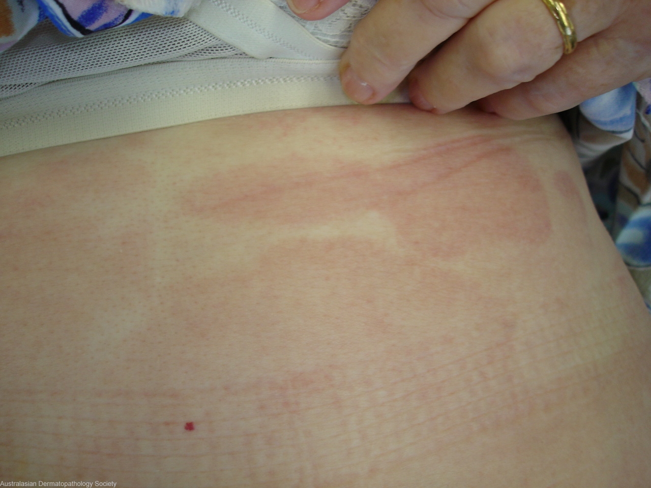
History: 2161ac Lady in her 60s with a 5 months history of fixed red patches that began in her groin and have spread over her abdomen. I suspect early T cell lymphoma of the skin DD early Morphoea. 3 biopsies from upper right abdominal patches
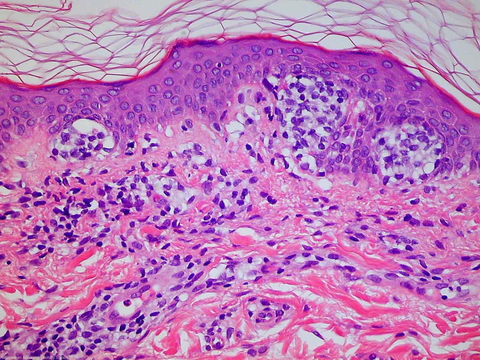
Description: Upper dermal atypical lymphocytes with epidermotropism
Comments: There is a moderately dense reasonably tight infiltrate of lymphocytes around upper dermal blood vessels. In addition some upper dermal interstitial lymphocytes are present. The lymphocytes show quite mild cytologic atypia. There is focal exocytosis of lymphocytes into the basal half of the epidermis with aggregation of these lymphocytes into small groups (Pautrier's micro-abscesses). The features present strongly suggest mycosis fungoides.