

Diagnosis: Extramammary Paget's
Description: Eroded infected surfaces of the vulva
Clinical Features: Erosions and fissures
Pathology/Site Features: Vulva
Sex: F
Age: 64
Submitted By: Ian McColl
Differential Diagnosis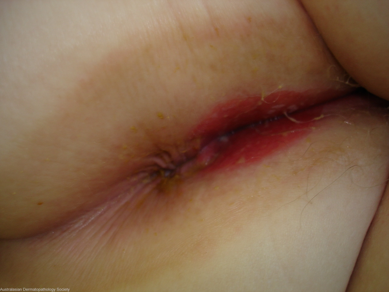
History: 2125bh Elderly female patient with a 3 year history of a slowly spreading perivulval and perivaginal rash,very painful,some adhesions,known Vin3. DD Pemphigoid, Extramammary Pagets, SCC in situ, Inflammatory lichen sclerosus Biopsies for histo and immunofluorescence .( Her original path diagnosis elsewhere of Vin3 was probably wrong)
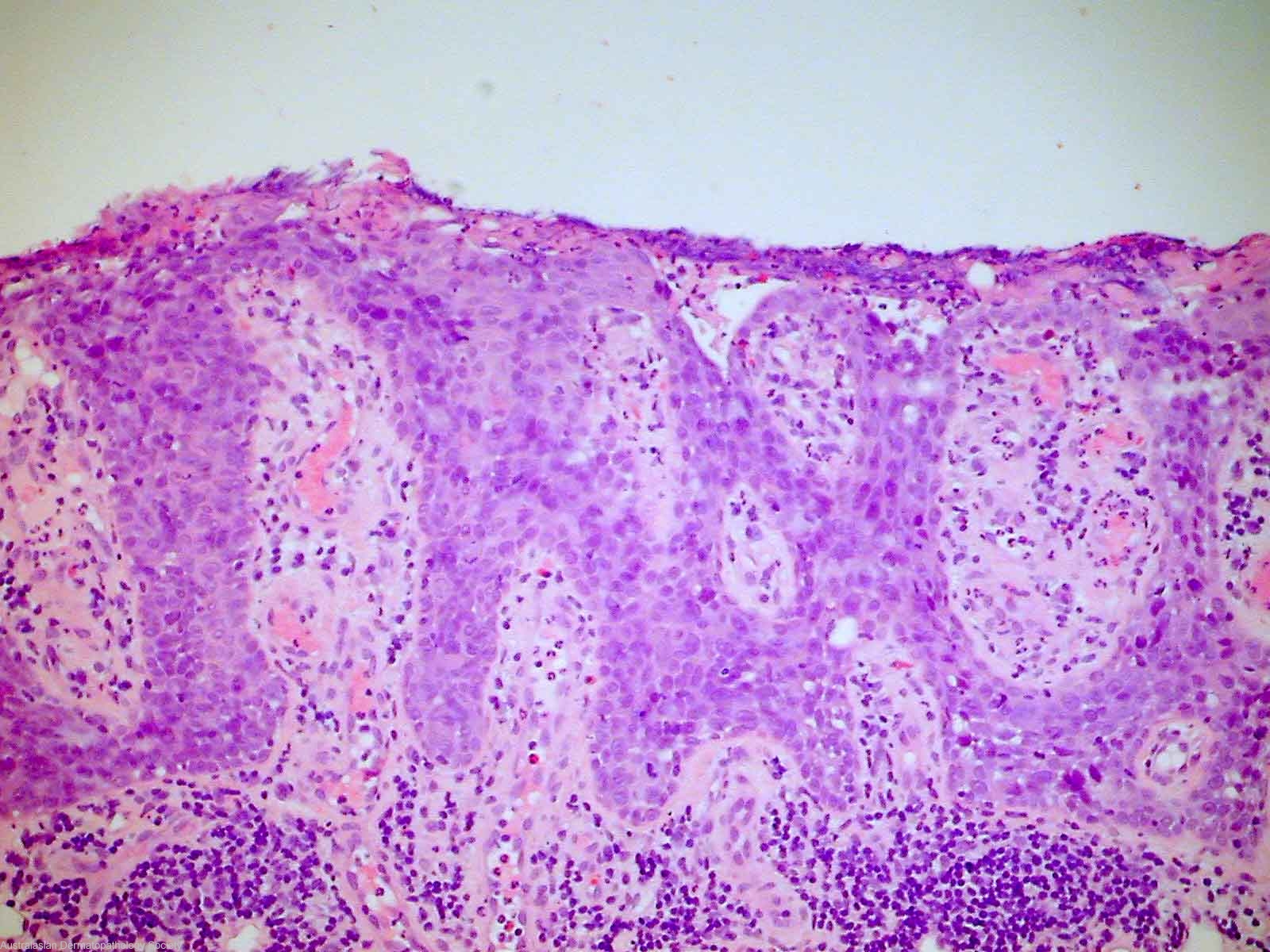
Description: Malignant cells fill the epidermis
Comments: The biopsy of perianal skin shows severely atypical cells within the epidermis with abundant cytoplasm , large nuclei and prominent nucleoli. The atypical cells extend to involve appendageal structures. There is no invasive malignancy. Immunohistochemical stains show the atypical cells to stain positively with cytokeratin 7 and epithelial membrane antigen. There is a negative reaction to S100, high molecular weight keratins and cytokeratin 20. The features are those of extra-mammary Paget's disease.