
Diagnosis: Tumid lupus
Description:
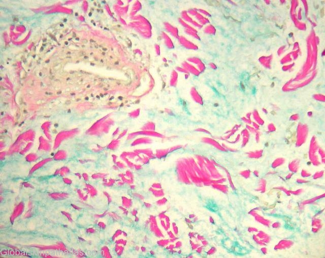
Clinical Features: Nodule
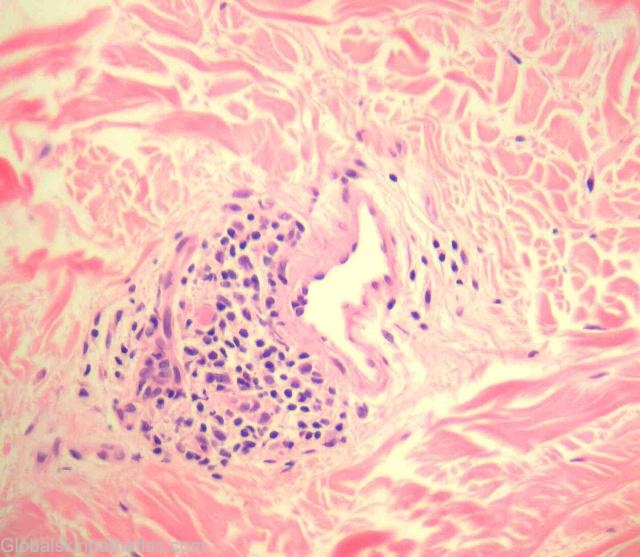
Pathology/Site Features: Perivascular infiltrate
Sex: M
Age: 43
Submitted By:
Differential Diagnosis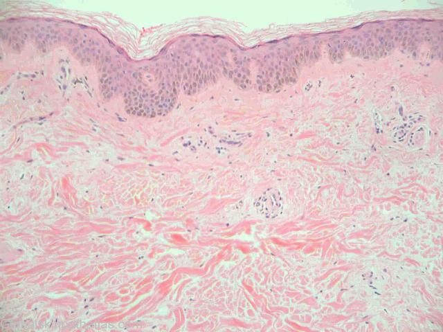
History:
Case submitted by Dr Trevor Beer Dermatopathologist Perth.
M 43, aboriginal. Asymptomatic nodules upper arm. The patient was hospitalised with lupus nephritis. During the admission his physician noted several nodules close to the axilla which were biopsied. They had not caused the patient any trouble or discomfort and he had no other cutaneous lesions.
Cutaneous lupus mucinosis is synonymous with tumid lupus / papulonodular mucinosis (1). It may be associated with either DLE or SLE and typically presents in males. Papules, nodules or plaques are mostly seen in the region of the head, neck and upper body. Some authors have suggested that it may represent a form of Jessner’s lymphocytic infiltrate.
The features in this case are characteristic with extensive dermal mucin and only a patchy dermal lymphocytic infiltrate. Epidermal changes of LE are not usually present.
The principal differentials are reticular erythematous mucinosis, scleredema and scleromyxedema as well as Jessner’s. Clinical correlation is the key to the correct diagnosis.
1. Kuhn. J Am Acad Derm 2003;48:901-8.