
Diagnosis: Pityriasis lichenoides
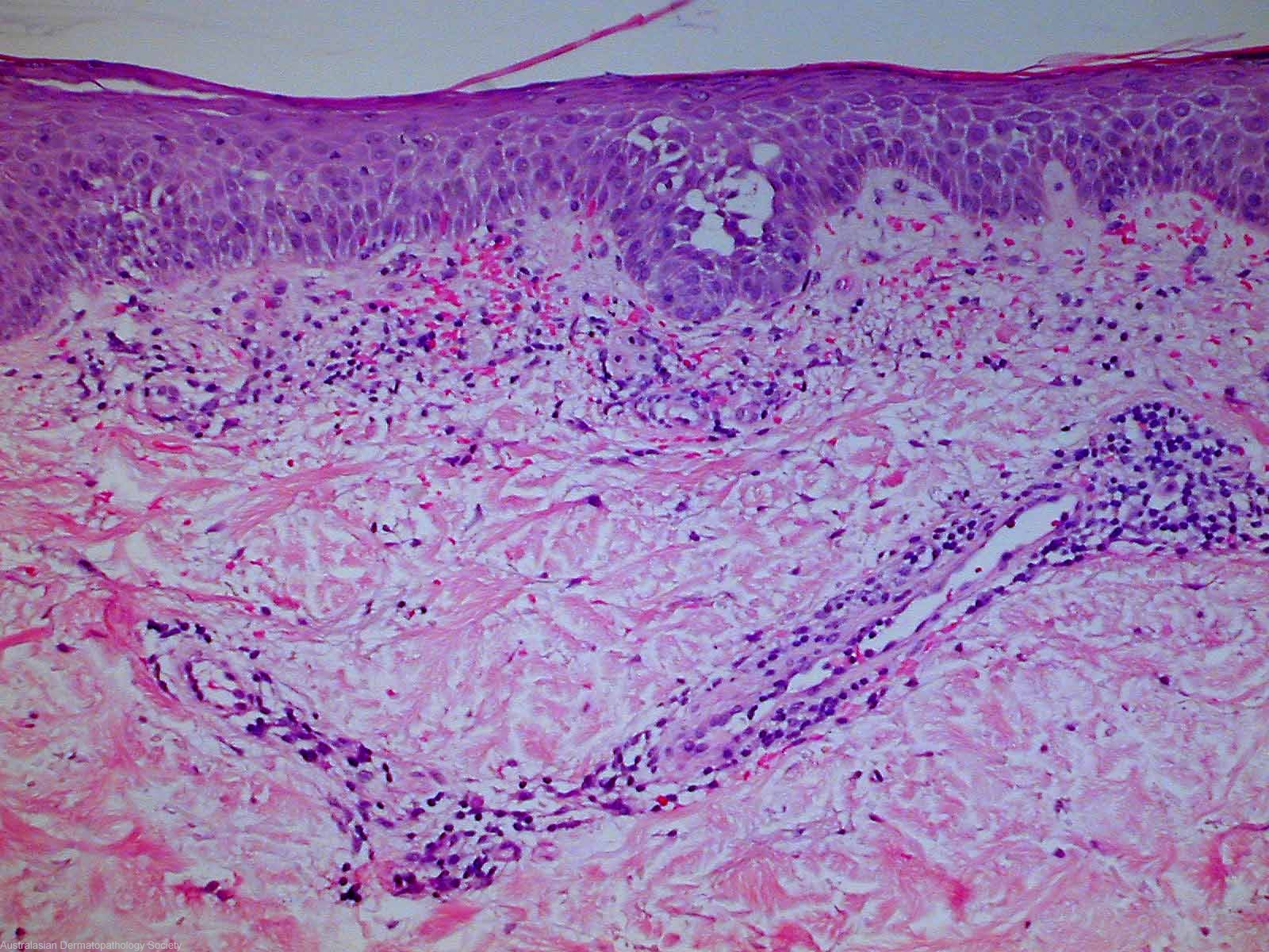
Description: mild acanthosis of the epidermis accompanied by moderate spongiosis. Focal lichenoid change involves the basal epidermis. There is a superficial dermal, quite tight perivascular infiltrate of lymphocytes. Red cell extravasation is prominent.
Clinical Features: Red,scaly
Pathology/Site Features: Basal layer degeneration
Sex: F
Age: 35
Submitted By: Ian McColl
Differential Diagnosis
History: 3627gh This lady has had an acral rash for two months that has failed to respond to topical steroids. It burns rather than itches. Clinically Pityriasis lichenoides. Biopsy done elsewhere suggested Pityriasis rosea which it clinically is not.
Description: Scaly papules leg
Comments: Sections show a biopsy of skin in which there is mild acanthosis of the epidermis accompanied by moderate spongiosis. Focal lichenoid change involves the basal epidermis. There is a superficial dermal, quite tight perivascular infiltrate of lymphocytes. Red cell extravasation is prominent. The perivascular inflammatory infiltrate also focally involves mid dermal vessels. There is focal exocytosis of lymphocytes into the spongiotic areas of the epidermis. No fungal organisms can be seen. The overall features would somewhat favour pityriasis lichenoides, however the presence of moderate spongiosis is quite unusual. Flegel's disease also a possibility/