
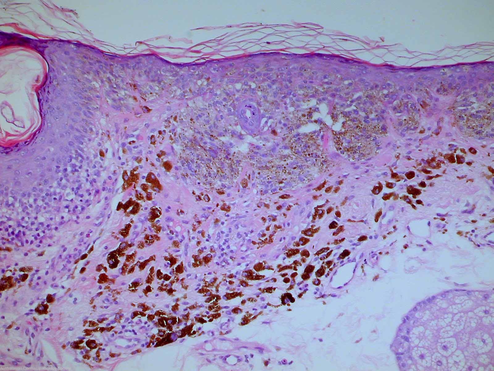
Diagnosis: Melanoma
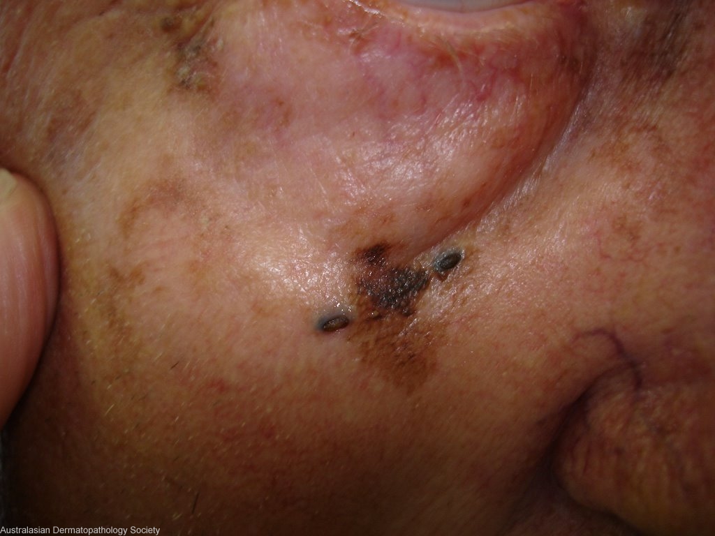
Description: Atypical facial pigmented lesion betweeen two comedones
Clinical Features: Macule black
Pathology/Site Features: Face
Sex: M
Age: 71
Submitted By: Ian McColl
Differential Diagnosis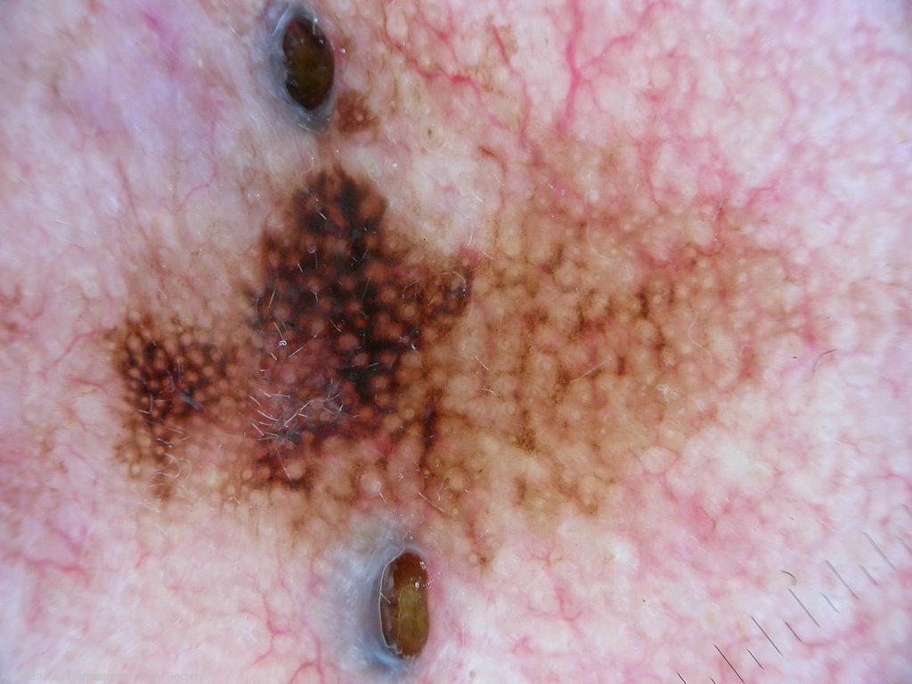
History: 2022dr This elderly man has noted this area of pigmentation develope over 6 months. The dermatoscope picture looks like lentigo maligna. Punch biopsy of darkest area. Appreciate full excisional biopsy better.
Description: Dermatoscope picture
Comments: Sections show a biopsy of skin in which there is a proliferation of atypical melanocytes restricted to the epidermis. They show both a nested and lentiginous pattern. Some large coalescing nests are present. There is upward epidermal spread of both single melanocytes and nests. The features best fit for a Level 1 superficial spreading malignant melanoma. The underlying dermis shows some fibrosis and prominent pigment incontinence.