
Diagnosis: Arthropod bite reaction
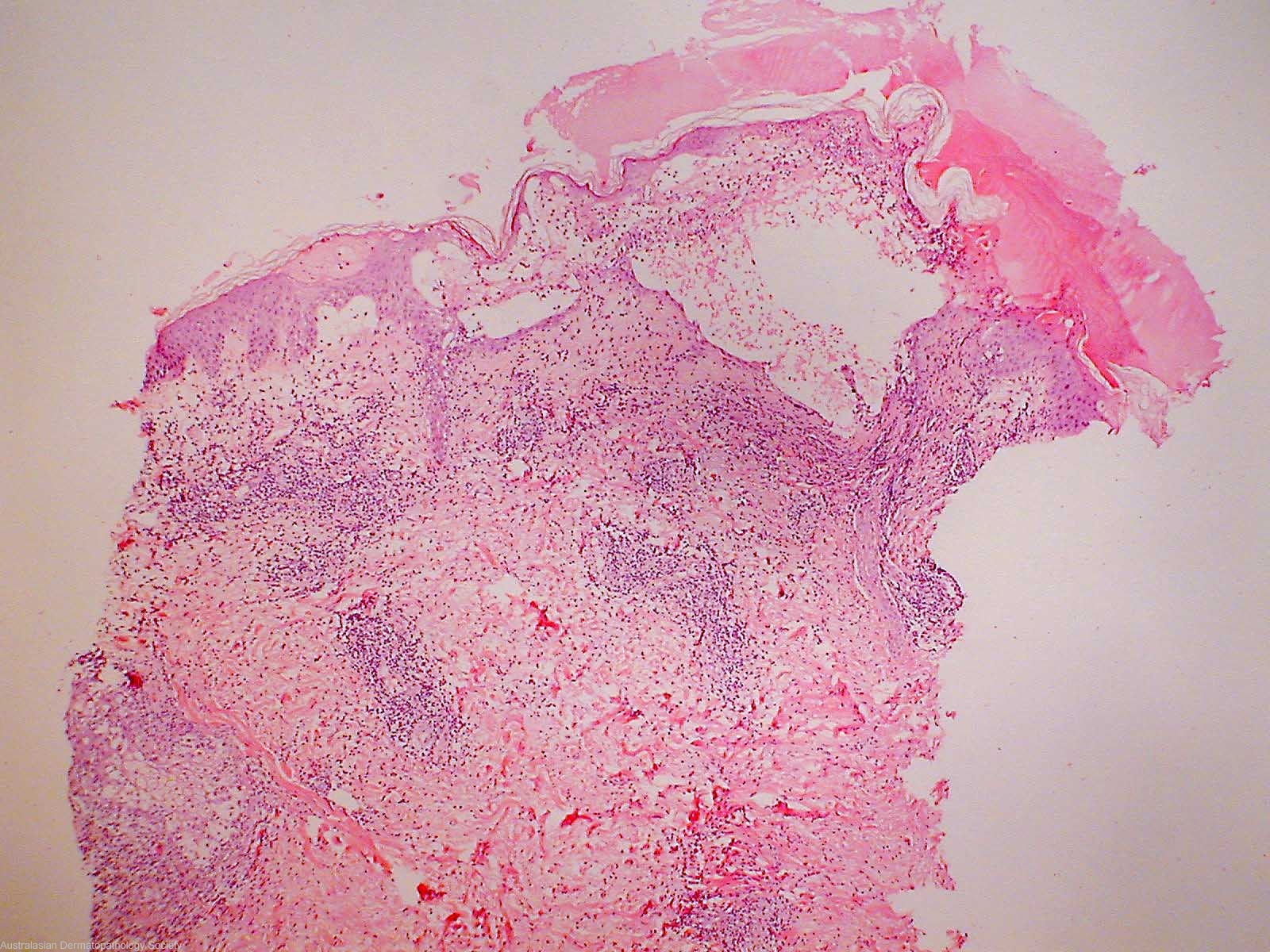
Description: There is some necrosis of the epidermis with intraepidermal blister formation. Both lymphocytes and eosinophils are present within the epidermis and the blister
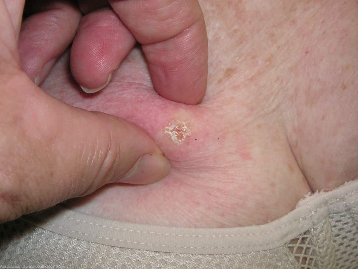
Clinical Features: Nodule pink
Pathology/Site Features: Eosinophils in the epidermis
Sex: F
Age: 68
Submitted By: Ian McColl
Differential DiagnosisHistory: 6896vf This lady has CLL which has been treated with chemotherapy. She developes these itchy firm nodules which have previously been reported as exaggerated insect bite reactions but they are occuring in non exposed areas (bedbugs excepted). Punch biopsy avoiding excoriated centre of lesion. See previous biopsy
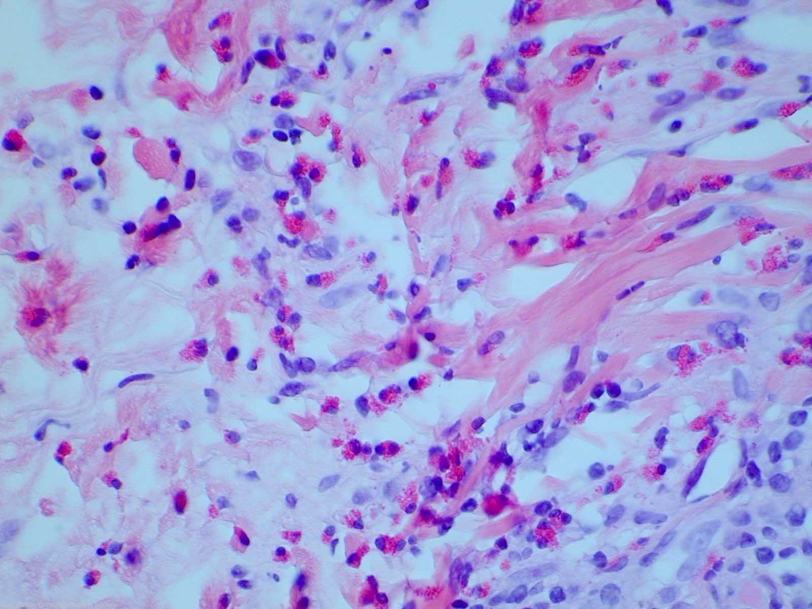
Description: Numerous eosinophils
Comments: Sections show a biopsy of skin in which there is a superficial mid and deep dermal infiltrate of lymphocytes and prominent eosinophils. The lymphocytes aggregate around blood vessels whilst the eosinophils are predominently interstitial in location. The infiltrate of both eosinophils and lymphocytes also extends into underlying subcutaneous fat. There is some necrosis of the epidermis with intraepidermal blister formation. Both lymphocytes and eosinophils are present within the epidermis and the blister.The histological changes in this biopsy are almost identical to the previous biopsy from this patient. In view of the absence of clinical evidence of arthropod bites, the term eosinophilic dermatosis of myeloproliferative disease is preferred. This terminology has recently been introduced to replace the previous terminology of exaggerated arthropod bite lesions associated with haematological disorders in cases where there is no evidence of bites.

