
Diagnosis: Melanoma
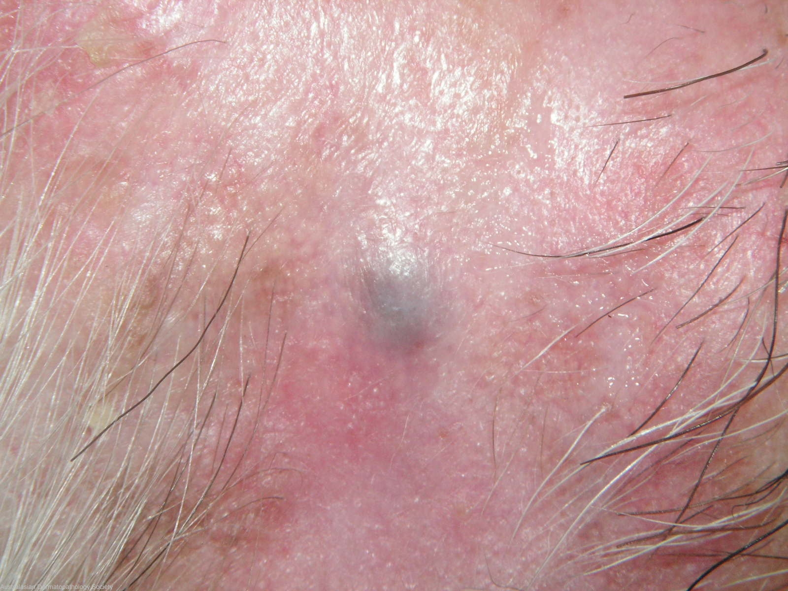
Description: Firm blue subcutaneous nodule
Clinical Features: Nodule,purple
Pathology/Site Features: Temple
Sex: M
Age: 81
Submitted By: Ian McColl
Differential Diagnosis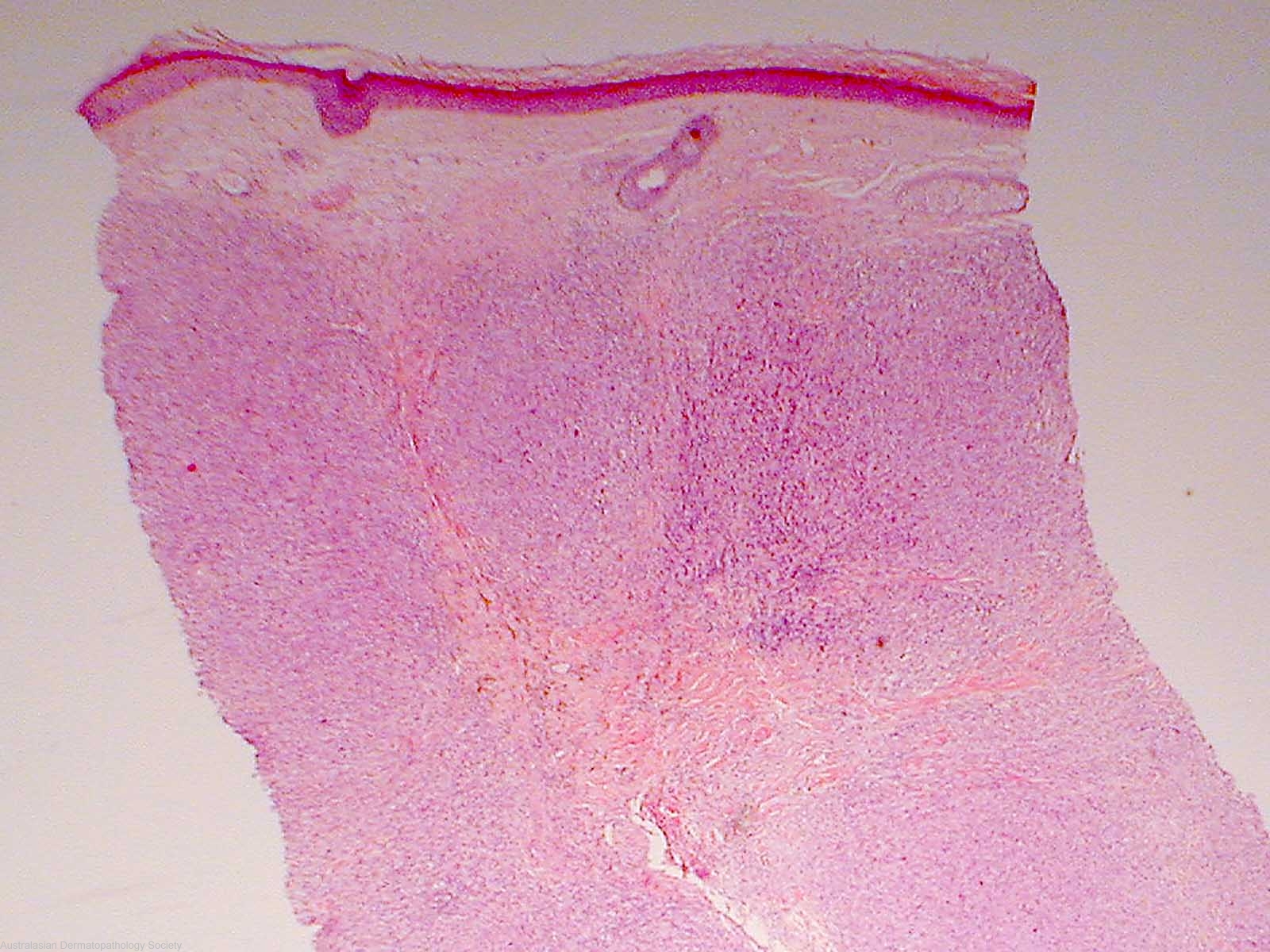
History: 2138hb This lesion has been present for three months on his temple. It is firm,not tender. The dermatoscope suggests a blue nevus but DD of hemorrhage into an eccrine hidrocystoma or thrombosed vein. Punch biopsy through the centre.
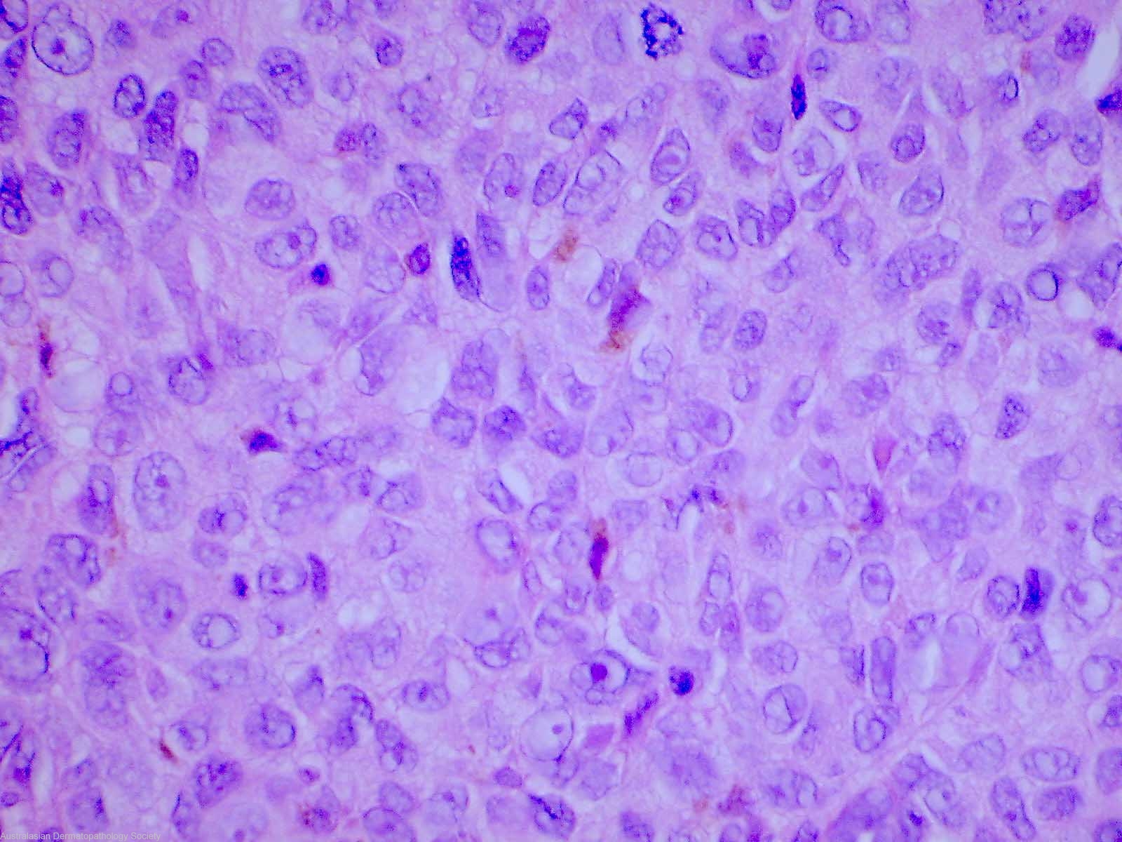
Description: Malignant melanoma.
Comments: Sections show a biopsy of skin that contains a deposit of malignant melanoma involving the mid and deep dermis. The upper dermis and epidermis are normal. The tumour consists of sheets of malignant cells with markedly pleomorphic nuclei. Some contain melanin pigment. The mitotic count is high. As there is no evidence of regression in the overlying epidermis in the biopsy, this dermal deposit of melanoma may represent a metastatic deposit. However, subsequent full excision showed a Level 5 nodular MM with evidence of regression of the epidermal component.