

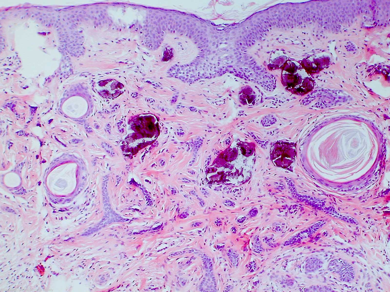
Diagnosis: Desmoplastic Trichoepithelioma
Description: White firm pearly lesion cheek
Clinical Features: Nodule,skin coloured
Pathology/Site Features: Cheek
Sex: M
Age: 26
Submitted By: Ian McColl
Differential Diagnosis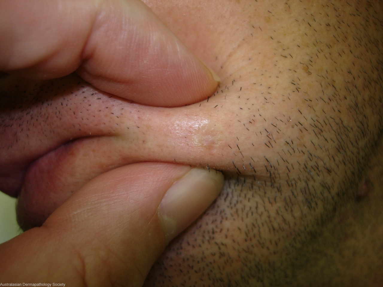
History: 1905tk Lesion on left and right cheeks for 9 months. Biopsy elsewhere reported as syringoma. Clinically desmoplastic trichoepithelioma but do you get desmoplastic syringomas or is that called microcystic adnexal carcinoma?
Description: Desmoplastic trichoepithelioma
Comments: Sections show a biopsy of a desmoplastic trichoepithelioma. There are strands of basaloid cells set within a desmoplastic stroma. There are small keratin cysts and some focal calcification.