
Diagnosis: Bowenoid papulosis
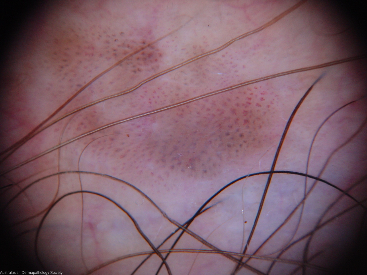
Description: Dermatoscopy
Clinical Features: Nodule,black
Pathology/Site Features: Penis
Sex: M
Age: 42
Submitted By: Ian McColl
Differential Diagnosis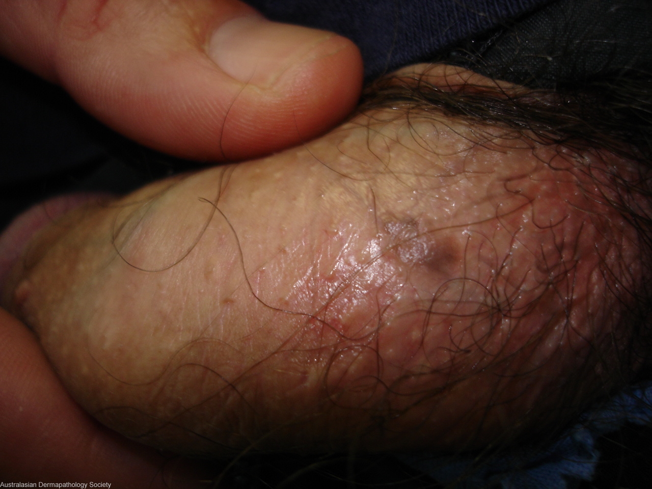
History:
5312pp This solitary pigmented lesion had been present on the shaft of the penis for 18 months. It was smoothe surfaced and slowly growing. DD nevus or seborrhoeic keratosis Shave biopsy
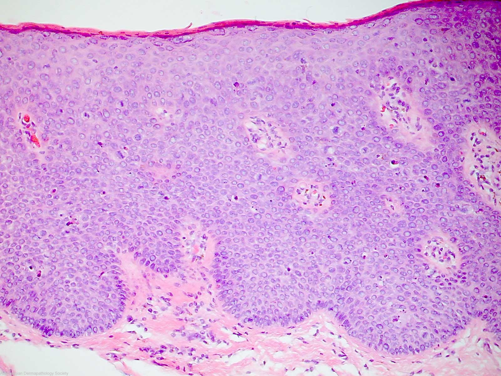
Description: bowenoid papulosis
Comments: Sections show a shaved area of bowenoid papulosis. There is epidermal hyperplasia with full thickness dysplasia. There are numerous mitoses throughout the epithelium and scattered dyskeratotic cells. There is a thin layer of overlying parakeratosis. There is no evidence of dermal invasion.