
Diagnosis: Melanoma
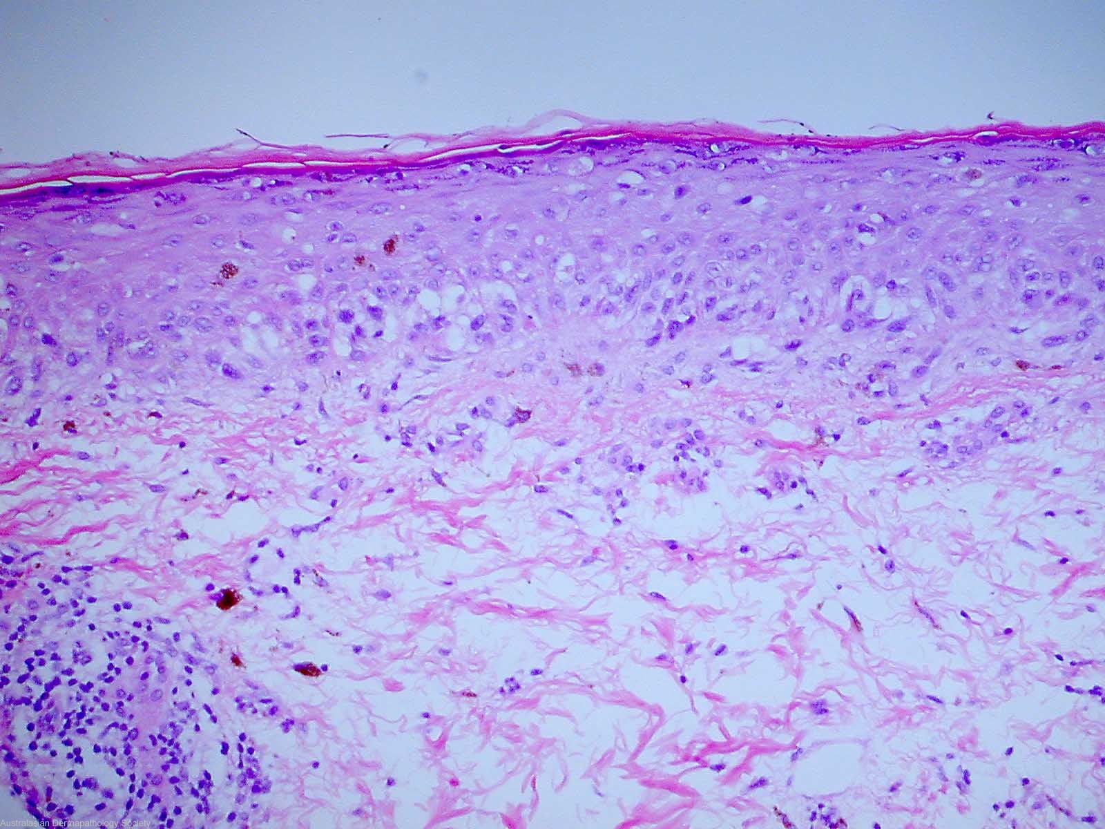
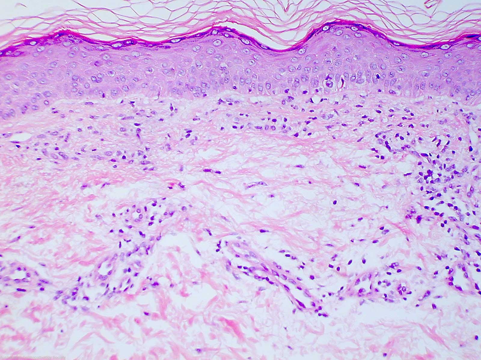
Description: It comprises a proliferation of atypical melanocytes forming irregular coalescing nests in the basal epidermis. There is focal upward epidermal spread
Clinical Features: Macule black
Pathology/Site Features: Melanoma nests
Sex: M
Age: 68
Submitted By: Ian McColl
Differential Diagnosis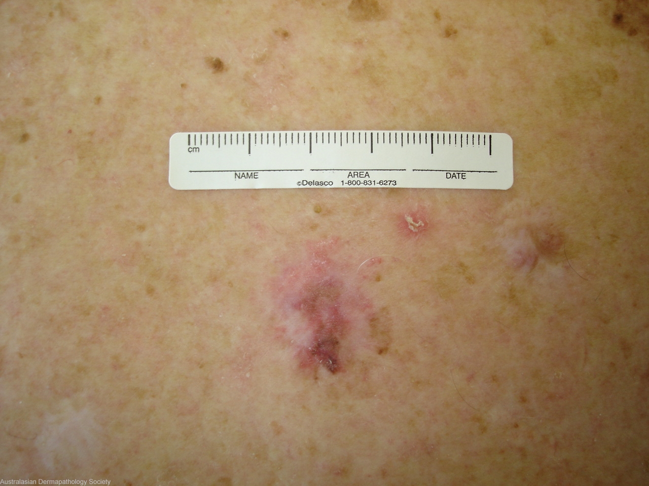
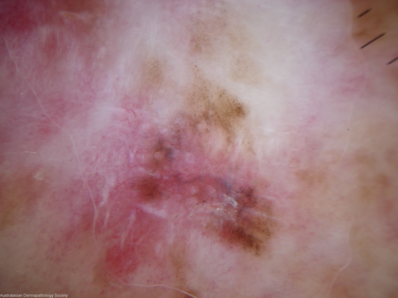
History: 4146ri This lesion on an elderly man's back looks suspicious with variable pigmentation and pink areas suggestive of regression. The lesion has not been treated before. Possible melanoma or other regressed pigmented lesion eg Dysplastic lentiginous junctional nevus of Kossard. Shave biopsy taken of the pigmented area at the base.
Description: Regression
Comments: Sections show a shave biopsy of a Level 1 superficial spreading malignant melanoma. It comprises a proliferation of atypical melanocytes forming irregular coalescing nests in the basal epidermis. There is focal upward epidermal spread. Some extension down hair follicle epithelium is also noted. There is no evidence of dermal invasion, however, there is evidence of regression.