

Diagnosis: Vasculitis
Description: Area of skin purpura and necrosis
Clinical Features: Purpura
Pathology/Site Features: Leg
Sex: F
Age: 56
Submitted By: Ian McColl
Differential Diagnosis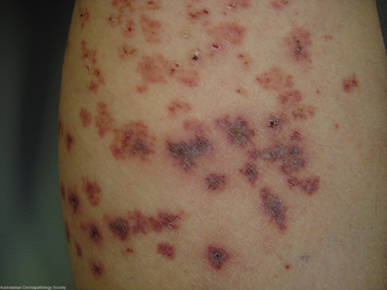
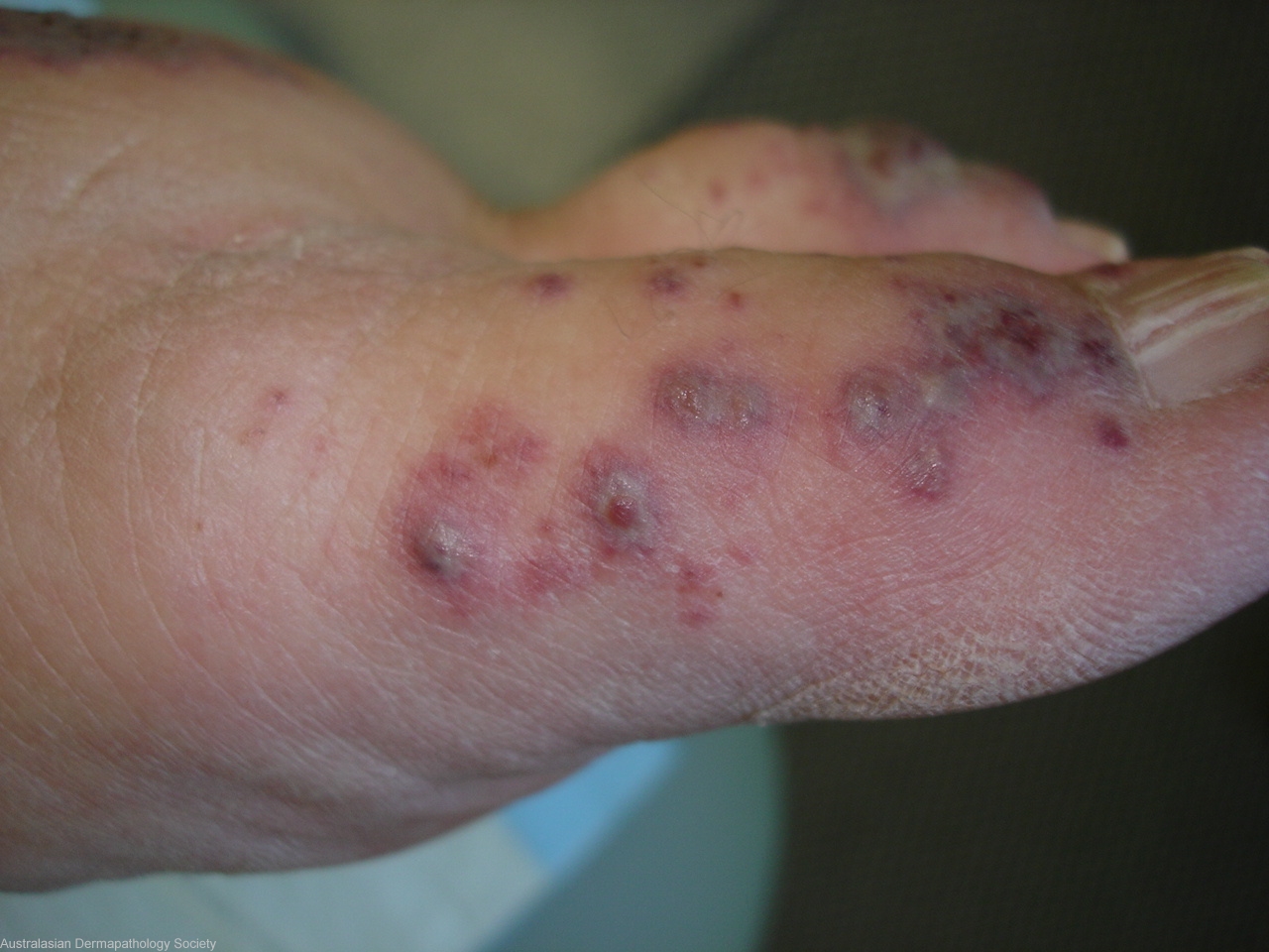
History: : This lady has a 3 week history of these lesions on her lower legs only from the knees down. She has a positive ana 1 in 640 anticentromere with a history of Raynauds in the winter. Has been on 50mgs oral steroids for 3 days when these pictures and biopsies were taken. Immunofluorescence ordered.
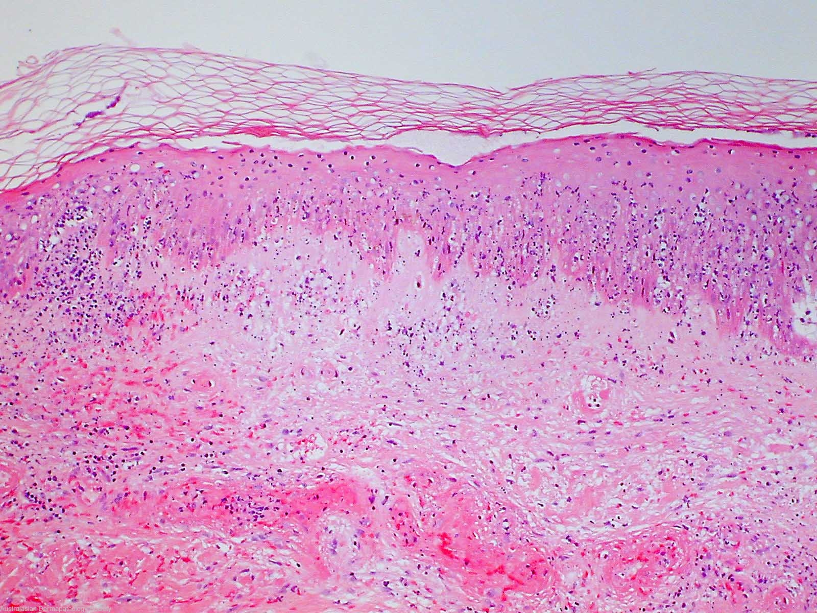
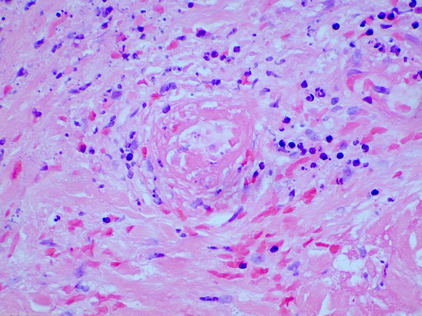
Description: Epidermal necrosis and underlying vasculiti
s
Comments: Sections show a biopsy of skin in which there is necrosis of the epidermis and upper dermis. Vessels throughout the full thickness of the dermis show features of vasculitis. There is fibrinoid deposition in the walls of the vessels with accumulation of neutrophils showing leucocytoclasis. Some of this vasculitis extends into underlying subcutaneous fat. There is a mild interstitial dermal infiltrate of neutrophils and some eosinophils. There are no histologic features that allow a specific aetiologic diagnosis for the vasculitis. Immunofluorescence microscopy appears negative for IgG, IgA, IgM, C3 and C1Q. Some fibrinogen is noted associated with dermal vessels.