

Diagnosis: Cellulitis
Description: Bullae and haemorrhage
Clinical Features: Blisters
Pathology/Site Features: Leg
Sex: M
Age: 45
Submitted By: Ian McColl
Differential Diagnosis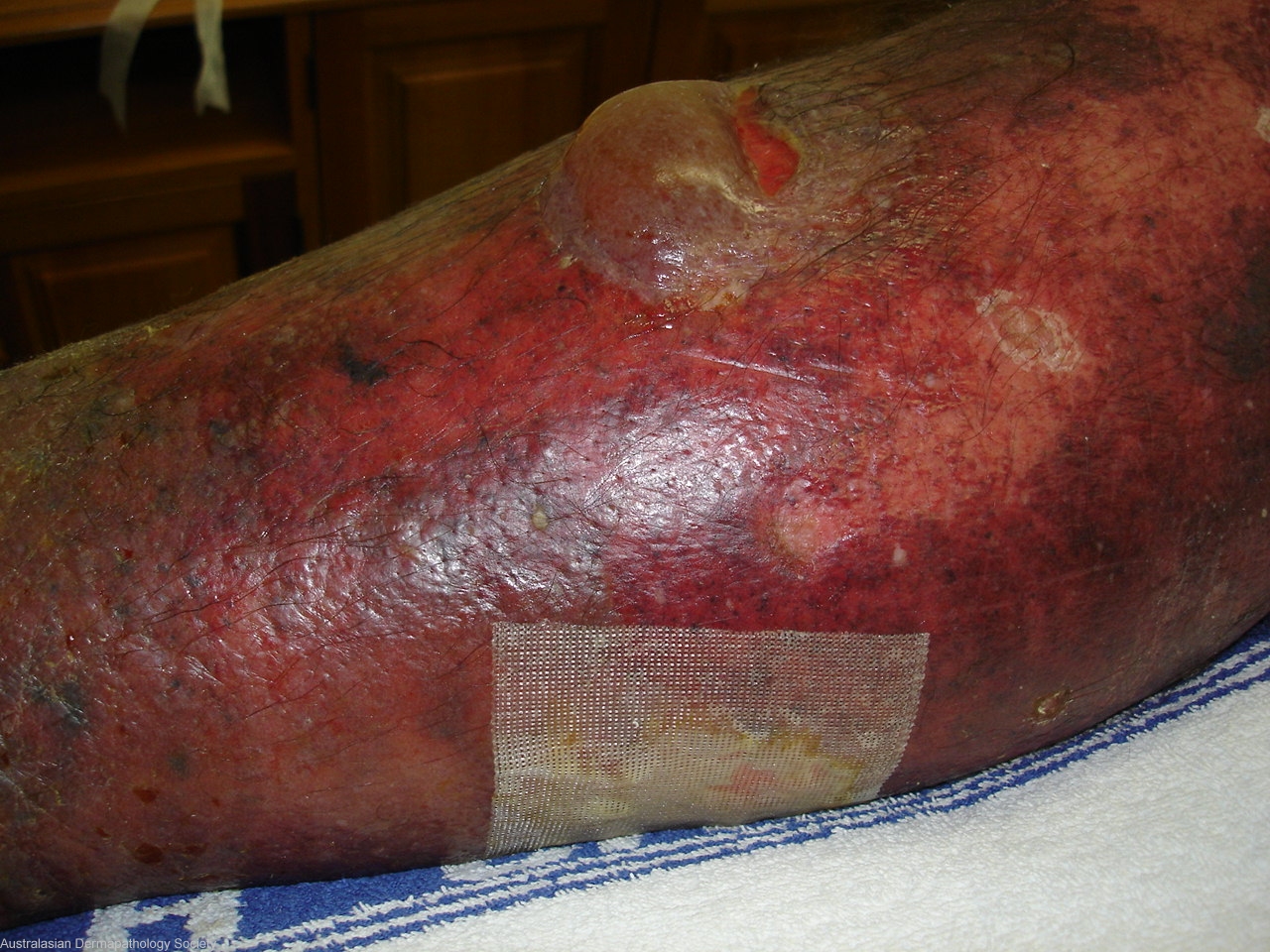
History: History: Sudden onset of painful swelling of the right leg with subsequent hemorrhage and bullae formation.Diagnosis Hemorrhagic cellulitis,query necrotizing fasciitis.He is an alcoholic with markedly abnormal lfts,diabetic and suffers from psoriasis.No history of injury.
Description: Bullae and skin hemorrhage
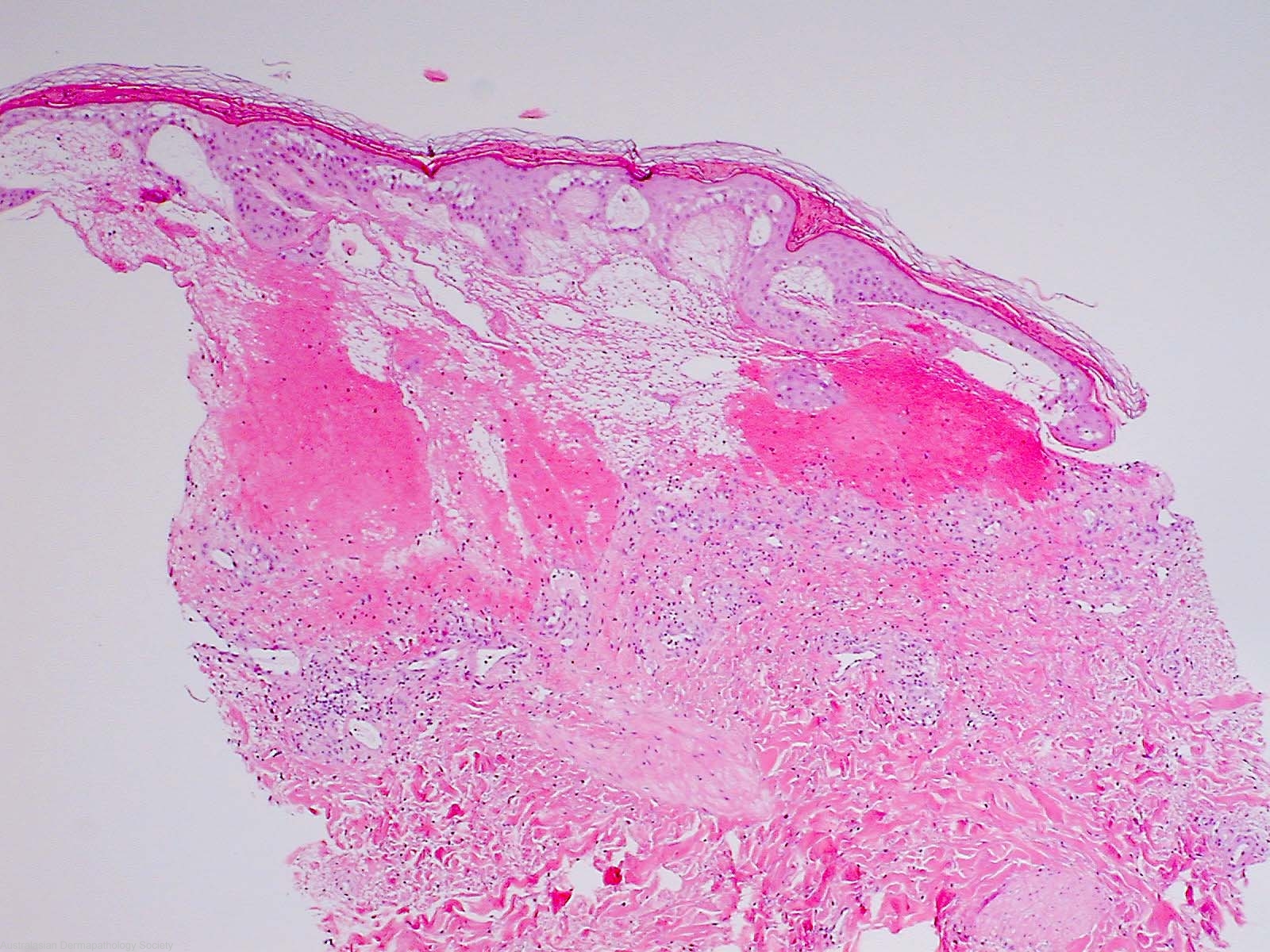
Comments: Sections show a biopsy of skin in which there is marked subepidermal oedema with vesicle formation. Haemorrhage is present within the vesicle. There is mild oedema of the rest of the dermis. This is accompanied by a mild infiltrate of neutrophils and lymphocytes. The infiltrate is heaviest in the upper dermis beneath the oedematous area. There is no evidence of dermal or epidermal necrosis. No bacteria can be seen on the Gram stain.The changes in all three biopsies are those of cellulitis. In the first two biopsies there is marked subepidermal oedema with bulla formation and subepidermal haemorrhage BULLOUS CELLULITIS WITH SUBEPIDERMAL HAEMORRHAGE